In vitro αξιολόγηση τριών τεχνικών προπαρασκευής κοιλότητας για ανάστροφη έμφραξη
|
|
|
- Στέφανος Δαμασκηνός
- 7 χρόνια πριν
- Προβολές:
Transcript

1 Αριστοτέλειο Πανεπιστήμιο Θεσσαλονίκης Τμήμα Οδοντιατρικής Εργαστήριο Ενδοδοντολογίας In vitro αξιολόγηση τριών τεχνικών προπαρασκευής κοιλότητας για ανάστροφη έμφραξη Μπελτές Χαράλαμπος Μεταπτυχιακός φοιτητής Διπλωματική διατριβή Θεσσαλονίκη 2006
2 Αριστοτέλειο Πανεπιστήμιο Θεσσαλονίκης Τμήμα Οδοντιατρικής Εργαστήριο Ενδοδοντολογίας In vitro αξιολόγηση τριών τεχνικών προπαρασκευής κοιλότητας για ανάστροφη έμφραξη Μπελτές Χαράλαμπος Μεταπτυχιακός φοιτητής Διπλωματική διατριβή Θεσσαλονίκη
3 Επιβλέπων μέλος Δ.Ε.Π.: Οικονομίδης Νικόλαος, Επίκουρος Καθηγητής 2
4 Περιεχόμενα Σελίδα 1. Περίληψη 4 2. Εισαγωγή Υλικό Μέθοδος Αποτελέσματα Συζήτηση Συμπεράσματα Πίνακες Βιβλιογραφία Εικόνες Ευχαριστίες
5 Περίληψη Η εισαγωγή των υπερήχων για την προπαρασκευή ανάστροφης κοιλότητας δημιούργησε νέα δεδομένα στην περιακρορριζική χειρουργική. Ο σκοπός της παρούσας ερευνητικής μελέτης είναι η in vitro αξιολόγηση τριών τεχνικών προπαρασκευής κοιλότητας για ανάστροφη έμφραξη. Χρησιμοποιήθηκαν 45 πρόσθια μονόριζα δόντια, άνω και κάτω γνάθου, ενδοδοντικά θεραπευμένα, τα οποία χωρίστηκαν σε τρεις ομάδες, των 15 δοντιών. Στην ομάδα Α οι προπαρασκευές πραγματοποιήθηκαν με στρογγύλη εγγλυφίδα Νο1 προσαρμοσμένη σε μικροχειρολαβή χαμηλών ταχυτήτων, στην ομάδα Β με ρύγχος υπερήχων ανοξείδωτου χάλυβα με διαμάντι στο κοπτικό άκρο και στην ομάδα Γ με ρύγχος υπερήχων ανοξείδωτου χάλυβα χωρίς διαμάντι στο κοπτικό άκρο. Η παρατήρηση έγινε με ηλεκτρονικό μικροσκόπιο σάρωσης (SEM) ύστερα από την δημιουργία αντιγράφων. Συγκρίθηκαν οι επιφάνειες των ακρορριζίων πριν και μετά την προπαρασκευή ανάστροφης κοιλότητας και αξιολογήθηκε ο αριθμός και το είδος των ρωγμών, η δημιουργία μικροανωμαλιών στα όρια των ανάστροφων κοιλοτήτων, καθώς και η επιφάνεια των εμφρακτικών υλικών μετά την εκτομή του ακρορριζικού τμήματος της ρίζας. Δεν βρέθηκαν στατιστικά σημαντικές διαφορές ανάμεσα στις τρεις τεχνικές, όσον αφορά τον αριθμό και το είδος των ρωγμών και των μικροανωμαλιών. Παρατηρήθηκαν μικροκενά ανάμεσα στα εμφρακτικά υλικά και τα τοιχώματα της οδοντίνης σε ορισμένα δείγματα. 4
6 Abstract The introduction of ultrasonics in root-end cavity preparation has brought new prospects in periapical surgery. The purpose of this study was the in vitro evaluation of three different root-end preparation techniques. 45 single-rooted anterior teeth, endodontically treated, were divided into three groups. Each group included 15 teeth. Group A was performed with a round bur No1 adapted in a slow-speed micro-handpiece, group B with a diamond-coated stainless steel ultrasonic tip and group C with a smooth stainless steel ultrasonic tip. Teeth were examined under scanning electron microscope (SEM) after replicas preparation of the specimens. The apical surfaces were compared before and after the rootend preparation. The number, the type of cracks and the marginal quality of the retrograde cavities were evaluated. The surface of root filling materials after rootend resection was also examined. There were no statistically significant differences in treatment results related to the number and the type of cracks, and the marginal chipping. Gaps were noted between the root filling materials and the walls of the root canal in few specimens. 5
7 Εισαγωγή Η συντηρητική ενδοδοντική θεραπεία θεωρείται η μέθοδος εκλογής για την αντιμετώπιση του μολυσμένου ριζικού σωλήνα, υπάρχουν όμως περιπτώσεις όπου κάτι τέτοιο δεν είναι εφικτό και η χειρουργική προσέγγιση κρίνεται αναγκαία. Λόγοι όπως, η πολυπλοκότητα του συστήματος του ριζικού σωλήνα, οι εμμένουσες περιακρορριζικές αλλοιώσεις, η παρουσία φυσικών φραγμών (ανατομικοί, ενασβεστιώσεις, προσθετικές αποκαταστάσεις, ενδορριζικοί άξονες, εμφρακτικά υλικά, σπασμένα μικροεργαλεία) δικαιολογούν ένα μικρό ποσοστό αποτυχίας της συντηρητικής ενδοδοντικής θεραπείας, που κυμαίνεται από 10-15%. (Gutmann 1991, Nair 1998) Σε περιπτώσεις που η επανάληψη της ενδοδοντικής θεραπείας δεν μπορεί να πραγματοποιηθεί ή δεν καταλήγει σε καλύτερα αποτελέσματα, η χειρουργική ενδοδοντική θεραπεία (περιακρορριζική χειρουργική) είναι η επόμενη εναλλακτική προσέγγιση. Γενικότερα, η διαδικασία περιλαμβάνει απομάκρυνση του κοκκιωματώδους ιστού, εκτομή του ακρορριζίου, προπαρασκευή και έμφραξη της ανάστροφης κοιλότητας. (Carr 1994) H προπαρασκευή ανάστροφης κοιλότητας και η τοποθέτηση εμφρακτικού υλικού έχει σκοπό να εκπληρώσει μία βασική βιολογική ανάγκη: την ερμητική έμφραξη κάθε ενεργού ή, ενδεχομένως, βλαπτικού παράγοντα εντός των φυσικών περιορισμών της ρίζας. Η δημιουργία κατάλληλης κοιλότητας σε σχήμα και μέγεθος που θα μπορεί, στην συνέχεια, να εμφραχθεί ερμητικά με κάποιο υλικό ανάστροφης έμφραξης, αποτελεί φραγμό έναντι των μικροοργανισμών και των προϊόντων τους που είτε έχουν παραμείνει στον ριζικό σωλήνα, είτε βρίσκονται στους περιακρορριζικούς ιστούς. Η μακροχρόνια αποφρακτική ικανότητα που εξασφαλίζει η σωστή προπαρασκευή και έμφραξη της ανάστροφης κοιλότητας πρέπει να ικανοποιεί αυτήν την βιολογική ανάγκη. Η ιδανική ανάστροφη κοιλότητα μπορεί να χαρακτηριστεί κοιλότητα ομάδας Ι, βάθους τουλάχιστον 3 mm μέσα στην οδοντίνη της ρίζας, με παράλληλα τοιχώματα που συμπίπτουν με την ανατομική μορφολογία του ριζικού σωλήνα. (Carr 1994, 1997) Περιορισμένα μέσα και τεχνικές έχουν χρησιμοποιηθεί για την προπαρασκευή ανάστροφης κοιλότητας. Η μοναδική τεχνική, τον τελευταίο αιώνα, ήταν η χρήση εγγλυφίδας προσαρμοσμένη στην κλασική χειρολαβή χαμηλών ή υψηλών ταχυτήτων με πολλά αρνητικά επακόλουθα λόγω, κυρίως, 6
8 του μεγάλου μεγέθους των χειρολαβών. Το 1939 ο Tangerud κατασκεύασε μια μικρογραφία της κλασικής χειρολαβής με κεφαλή ύψους 2,5 mm και εύρους 4 mm, (Κavo Ltd., UBECO, DynaDent) για να διευκολυνθεί η πρόσβαση και η προπαρασκευή ανάστροφης κοιλότητας στο ακρορριζικό τμήμα της ρίζας. Στην μικροχειρολαβή προσαρμόζεται κυλινδρική εγγλυφίδα Νο1 ή ανεστραμμένου κώνου Νο34. Αυτή η τεχνική θεωρήθηκε η πιο καθιερωμένη στην κλινική πράξη μέχρι την εισαγωγή των υπερήχων στην χειρουργική ενδοδοντία.(tangerud 1993) Πρώτες αναφορές στην βιβλιογραφία σαν μεμονωμένες κλινικές περιπτώσεις γίνονται από τον Richman (1957), από τους Bertrand και συν. (1976) και από τους Flath και Hicks (1987). Ειδικά σχεδιασμένα ρύγχη για συσκευές υπερήχων σχεδιάστηκαν και κατασκευάστηκαν για πρώτη φορά το 1990 (Excellence in Endodontics, San Diego, CA, USA) (Carr 1990, 1992, Pannkuk 1992). Μέχρι σήμερα έχουν κατασκευαστεί από διάφορες εταιρίες αρκετά είδη, με τροποποιήσεις ως προς το σχήμα, το μέγεθος, την γωνία, την συχνότητα δόνησης και το υλικό του κοπτικού άκρου. Ως προς την συχνότητα δόνησης διαδεδομένα είναι τα ρύγχη υπερήχων, ενώ έχουν δοκιμαστεί και ρύγχη χαμηλότερης ηχητικής συχνότητας. Ως προς το υλικό του κοπτικού άκρου υπάρχουν ρύγχη υπερήχων ανοξείδωτου χάλυβα με ή χωρίς διαμάντι στο κοπτικό άκρο, ενώ πρόσφατα κυκλοφόρησαν στο εμπόριο και ρύγχη ανοξείδωτου χάλυβα με νιτρικό ζιρκόνιο στο κοπτικό άκρο (ΚiS/Spartan-Obtura, Fectron,MI). Η χρήση των υπερήχων στην χειρουργική ενδοδοντία έχει γίνει ευρέως αποδεκτή καθώς τα πλεονεκτήματα σε σχέση με την κλασική μέθοδο είναι πολλά. Οι μικρότερες διαστάσεις τους, η ευκολότερη πρόσβαση στην περιοχή του ακρορριζικού τμήματος της ρίζας σε συνδυασμό με την μειωμένη απομάκρυνση οστίτη ιστού, η ελαχιστοποίηση ή ο μηδενισμός της γωνίας εκτομής (εκτομή κάθετη ως προς τον επιμήκη άξονα του δοντιού) και η συνολικά μικρότερη έκθεση οδοντινοσωληναρίων είναι μερικά από αυτά. Σε σχέση με την διαμόρφωση της ανάστροφης κοιλότητας μπορούν να παρασκευαστούν μικρότερες και πιο βαθιές κοιλότητες, με πιο καθαρά τοιχώματα (απομάκρυνση οδοντικού τρίματος- debris), με καλύτερο σχήμα συγκράτησης και πιο ευθυγραμμισμένες σε σχέση με τον επιμήκη άξονα του ριζικού σωλήνα. (Carr 1997, Wuchenich 1994, Gutmann 1994, Gorman 1995) Η προπαρασκευή με ρύγχη υπερήχων έναντι της κλασικής μεθόδου παρέχει πλεονεκτήματα στην 7
9 διαμόρφωση και στον καθαρισμό ριζών με βαθιές αύλακες και παρουσία ισθμών, μειώνοντας τις πιθανότητες διάτρησης της ρίζας. (Engel 1995, Zuolo 1999) Όμως παρά τα πλεονεκτήματα της χρήσης των υπερήχων στην περιακρορριζική χειρουργική, οι Saunders και συν. (1994),σε in vitro μελέτη τους, ήταν οι πρώτοι που ανέφεραν ότι η δημιουργία ρωγμών στην επιφάνεια του ακρορριζικού τμήματος της ρίζας μετά την προπαρασκευή ανάστροφης κοιλότητας με ρύγχη υπερήχων παρατηρείται συχνότερα, σε σχέση με την προπαρασκευή με στρογγύλη εγγλυφίδα προσαρμοσμένη σε μικροχειρολαβή χαμηλών στροφών. Ο Abedi και συν. (1995) βρήκαν ευρήματα παρόμοια με αυτά των Saunders και συν. Τα αποτελέσματα άλλων μελετών έδειξαν μεγαλύτερο ποσοστό ρωγμών όταν οι τα ρύγχη υπερήχων χρησιμοποιούνται σε υψηλές συχνότητες παρά σε μέσες και χαμηλές συχνότητες (Frank 1996, Layton 1996), ενώ ο Gray και συν. (2000) δεν βρήκαν σημαντικές διαφορές σε προπαρασκευές με υψηλές, μέσες και χαμηλές συχνότητες. Σε αντίθεση με τις προηγούμενες αναφορές, αποτελέσματα άλλων μελετών δεν έδειξαν στατιστικά σημαντικές διαφορές μεταξύ προπαρασκευών με υπέρηχους και την κλασική τεχνική. Μικρότερος αριθμός ή και καμία ρωγμή παρατηρήθηκε όταν οι προπαρασκευές έγιναν με ρύγχη υπερήχων. (Lloyd 1996, Waplington 1997, Lin 1998, Navarre 2002) Συγκριτικές μελέτες έχουν γίνει και μεταξύ των διαφόρων τύπων υπερήχων χωρίς να παρατηρηθούν στατιστικά σημαντικές διαφορές. (Zuolo 1999, Brent 1999, Sultan 1995) Στην προσπάθεια να αποδοθούν σε πιο αξιόπιστο βαθμό οι κλινικές συνθήκες, έχουν γίνει έρευνες σε ανθρώπινα πτώματα και τα ευρήματα έδειξαν λιγότερες ρωγμές στην επιφάνεια του ακρορριζίου, σε σύγκριση με έρευνες στις οποίες χρησιμοποιήθηκαν εξαγχθέντα δόντια. (Gray 2000, Min 1997, Calzonetti 1998, De Bryne & De Moor 2005) Επίσης, σαν συνέπεια της χρήσης υπερήχων, έχουν αξιολογηθεί, μικροανωμαλίες στα όρια της ανάστροφης κοιλότητας (Lloyd 1996, Waplington 1997, Gray 2000, Gondim 2002, De Bryne & De Moor 2005) Ο σκοπός της παρούσας μελέτης είναι η αξιολόγηση της επίδρασης πάνω στην επιφάνεια του ακρορριζίου δύο διαφορετικών τύπων υπερήχων και της κλασσικής τεχνικής, ύστερα από την προπαρασκευή κοιλότητας για ανάστροφη έμφραξη. Η σύγκριση έγινε ανάμεσα σε ρύγχος υπερήχων ανοξείδωτου χάλυβα με διαμάντι στο κοπτικό άκρο, σε ρύγχος υπερήχων 8
10 ανοξείδωτου χάλυβα χωρίς διαμάντι στο κοπτικό άκρο και στρογγύλη εγγλυφίδα Νο1 προσαρμοσμένη σε μικροχειρολαβή χαμηλών ταχυτήτων. Αξιολογήθηκε, ο αριθμός και το είδος των ρωγμών πριν και μετά την προπαρασκευή της ανάστροφης κοιλότητας, η δημιουργία μικροανωμαλιών στα όρια των ανάστροφων κοιλοτήτων, καθώς και η επιφάνεια των εμφρακτικών υλικών μετά την εκτομή του ακρορριζικού τμήματος της ρίζας. 9
11 Υλικό Μέθοδος Χρησιμοποιήθηκαν 45 πρόσθια μονόριζα δόντια, προσφάτως εξαγχθέντα, άνω και κάτω γνάθου. Η επιλογή τους έγινε με κριτήρια την ευθεία πορεία της ρίζας και τον ένα ριζικό σωλήνα σε κάθε δόντι, με την βοήθεια λήψης ψηφιακής ακτινογραφίας. Δόντια με έντονες κάμψεις και περισσότερους από έναν ριζικό σωλήνα απορρίφθηκαν. Ύστερα από την εξαγωγή των δοντιών τα υπολείμματα και οι μαλακοί ιστοί που παρέμειναν στην επιφάνεια της ρίζας απομακρύνθηκαν με την βοήθεια αποστειρωμένης γάζας. Τα δόντια τοποθετήθηκαν σε διάλυμα υποχλωριώδους νατρίου 2,5% για 30 λεπτά και στη συνέχεια διατηρήθηκαν καθ όλη την διάρκεια του πειράματος μέσα σε αποσταγμένο νερό. Συντηρητική ενδοδοντική θεραπεία Ακολούθησε εκτομή του μυλικού τμήματος των δοντιών στο ύψος της αδαμαντινοοστεϊνικής ένωσης με κυλινδρικό διαμάντι (Komet, , GmbH&Co., Ger.) σε υψηλές ταχύτητες, για διευκόλυνση της προπαρασκευής των ριζικών σωλήνων και για καλύτερη τυποποίηση του μήκους τους. Ύστερα από ανίχνευση και εύρεση των μυλικών στομίων ορίστηκε το μήκος εργασίας με την έξοδο ενός μικροεργαλείου, ρίνης Κ Νο10, από το ακρορριζικό τρήμα και αφαίρεση 1mm από το σημείο αυτό. Η προπαρασκευή των ριζικών σωλήνων έγινε με την χρήση μηχανοκίνητων περιστρεφόμενων μικροεργαλείων Ni-Ti του συστήματος Protaper (Densply, Maillefer, Switz.). Τα μικροεργαλεία χρησιμοποιήθηκαν με βάση το πρωτόκολλο που προτείνεται από τον κατασκευαστή, με την ακόλουθη σειρά: Sx-S1-S2-F1-F2-F3. Οι διακλυσμοί κατά τις προπαρασκευές των ριζικών σωλήνων έγιναν με διάλυμα υποχλωριώδους νατρίου 2,5%, ενώ χρησιμοποιήθηκε λιπαντικός παράγοντας (Glyde TM ) κατά την χρήση των μηχανοκίνητων μικροεργαλείων και τελικός διακλυσμός με 5ml αποσταγμένο νερό. Οι ριζικοί σωλήνες στεγνώθηκαν με κώνους χάρτου μεγέθους Medium (Roeko, Coltene, Ger.) και τοποθετήθηκαν τυποποιημένοι κώνοι γουταπέρκας του συστήματος Protaper (Dentsply,Maillefer, Switz.) έτσι ώστε να δημιουργηθεί το αίσθημα της ενσφήνωσης στο ακρορρίζιο (tug back). Χρησιμοποιήθηκε φύραμα εποξυλικής ρητίνης (2Seal, VDW,GmbH, Ger.) και ακολουθήθηκε η τεχνική έμφραξης της κάθετης συμπύκνωσης μέσω 10
12 του συστήματος συνεχόμενου θερμού κύματος - System B. Όπου κρίθηκε απαραίτητη η χρησιμοποίηση και δευτερευόντων κώνων γουταπέρκας, αυτό έγινε με κώνους μη τυποποιημένους, μεγέθους Fine Medium και Fine Fine (Autofit GT; SybronEndo, CA, USA), οι οποίοι συμπυκνώθηκαν με πλάγιο συμπυκνωτήρα RCSD T11 (Hu-Friedy,USA). Η κάθετη συμπύκνωση έγινε με συνδυασμό του συστήματος System B (ρύγχη μεγέθους Μedium, Medium Fine) και με κάθετους συμπυκνωτήρες Νο2 και Νο3 (Dentsply, Maillefer, Switz.). (Buchanan 2004) Τα μυλικά στόμια αποφράχθηκαν με την τοποθέτηση υαλοϊονομερούς τροποποιημένης ρητίνης (Ιonosit TM ) και τα δόντια παρέμειναν για τουλάχιστον 72 ώρες σε υγρό περιβάλλον χωρίς να ασκηθεί καμία άλλη ενέργεια, για πλήρη πήξη του φυράματος. Όλα τα δόντια εξετάστηκαν για ύπαρξη τυχόν επιμηκών καταγμάτων κατά την διάρκεια αυτού του σταδίου μέσω οπτικού μικροσκοπίου σε μεγέθυνση x5. Σε ένα δόντι παρατηρήθηκε κάταγμα κατά μήκος της ρίζας μετά την έμφραξη του ριζικού σωλήνα και αντικαταστάθηκε. Εκτομή του ακρορριζίου Η εκτομή του ακρορριζικού τμήματος έγινε σε απόσταση 3mm από το ανατομικό ακρορρίζιο, κάθετα ως προς τον επιμήκη άξονα της ρίζας. Χρησιμοποιήθηκε δίσκος κοπής από διαμάντι 0,3mm τοποθετημένος σε μικροτόμο σκληρών ιστών (Isomet, Buehler, Evanston) με ταχύτητα στροφών και συνεχή καταιονισμό νερού. Στην συνέχεια έγινε αρχικός έλεγχος των επιφανειών των ριζών μετά την ακρορριζεκτομή με την χρήση στερεομικροσκοπίου σε μεγέθυνση x12 (Carl Zeiss/ stemi DV4). Οι επιφάνειες φωτογραφήθηκαν με την χρήση ψηφιακής φωτογραφικής μηχανής (Sony DSC-S85) προσαρμοσμένη στο στερεομικροσκόπιο και αξιολογήθηκαν για την ύπαρξη αρχικών ρωγμών με παρατήρηση των εικόνων σε ηλεκτρονικό υπολογιστή (λογισμικό πρόγραμμα επεξεργασίας εικόνας, Photoshop CS ed.) Τα δόντια ξεπλύθηκαν με τρεχούμενο αποσταγμένο νερό για 3 λεπτά και τοποθετήθηκαν σε διάλυμα ΕDTA 17% για 1 λεπτό. Η απομάκρυνση του ΕDTA έγινε με εκ νέου τοποθέτηση σε αποσταγμένο νερό για άλλα 5 λεπτά και τα δόντια στεγνώθηκαν με την χρήση αεροσύριγγας. 11
13 Λήψη αρχικών αποτυπωμάτων Η λήψη αρχικών αποτυπωμάτων των επιφανειών των ακρορριζίων έγινε με την χρήση υψηλής ευκρίνειας λεπτόρευστης σιλικόνης (R-Siline Light, GmbH, Dh.). Η έγχυση έγινε με ειδικά ρύγχη της ίδιας εταιρείας πάνω στις επιφάνειες των ακρορριζίων και ως μίτρες χρησιμοποιήθηκαν πλαστικά καπάκια διαστάσεων 0,5x0,5cm περίπου. Μετά από 5 λεπτά τα αποτυπώματα διαχωρίστηκαν από τις επιφάνειες των δοντιών και ελέγχθηκαν για τυχόν ατέλειες μέσω οπτικού μικροσκοπίου σε μεγέθυνση x10. Σε περιπτώσεις ατελειών πραγματοποιήθηκε νέα λήψη αποτυπωμάτων. Τεχνική αντιγραφής (replicas) Η τεχνική αντιγραφής έγινε με την επακόλουθη πλήρωση των κυπελλοειδούς σχήματος αποτυπωμάτων με χαμηλού ιξώδους εποξυλική ρητίνη (Epoxy Embedding Medium, Fluka, Sigma-Aldrich Chemie GmbH, Ger.), με βάση τις οδηγίες του κατασκευαστή. Οι μίτρες στην συνέχεια τοποθετήθηκαν σε κλίβανο σταθερής θερμοκρασίας 60 βαθμών Κελσίου για 24 ώρες για να διασφαλιστεί ο πλήρης πολυμερισμός της ρητίνης, πάντα με βάση τις οδηγίες του κατασκευαστή. (Εικόνα Ι ) Προπαρασκευή ανάστροφων κοιλοτήτων Τα δόντια χωρίστηκαν σε τρεις ομάδες (Α, Β, Γ) με βάση την τεχνική προπαρασκευής που ακολουθήθηκε. Ομάδα Α: Περιελάμβανε 15 δόντια που προπαρασκευάστηκαν με την κλασσική τεχνική. Χρησιμοποιήθηκε στρογγύλη εγγλυφίδα Νο1 (Komet, ISO500, GmbH&Co., Ger.) προσαρμοσμένη σε μικροχειρολαβή χαμηλών ταχυτήτων (KAVO, Biberach,Ger. / στροφές ανά λεπτό). Ομάδα Β: Περιελάμβανε 15 δόντια που προπαρασκευάστηκαν με την τεχνική υπερήχων. Χρησιμοποιήθηκε ρύγχος υπερήχων ανοξείδωτου χάλυβα CT-1 με διαμάντι στο κοπτικό άκρο (SybronEndο,Orange,USA) προσαρμοσμένο σε συσκευή υπερήχων, σε συχνότητα 30 khz ( μέση ισχύς 7,πρόγραμμα S) (Suni-Max, Satelec, Merignac, Cedex, Fr.). Ομάδα Γ: Περιελάμβανε 15 δόντια που προπαρασκευάστηκαν με την τεχνική υπερήχων. Χρησιμοποιήθηκε ρύγχος υπερήχων ανοξείδωτου χάλυβα CT-1 χωρίς διαμάντι στο κοπτικό άκρο (SybronEndo,Orange,USA) 12
14 προσαρμοσμένο σε συσκευή υπερήχων, σε συχνότητα 30 khz (μέση ισχύς 7,πρόγραμμα S) (Suni-Max, Satelec, Merignac, Cedex, Fr.). Όλες οι προπαρασκευές έγιναν από τον ίδιο επεμβαίνοντα με φορά προς την μυλική κατεύθυνση της ρίζας και συνεχή καταιονισμό νερού. Η κίνηση ήταν συνεχόμενη, «εμπρός πίσω», με ελάχιστη πίεση ( feather-like movement ). (De Bryne & De Moor 2005) Στην ομάδα Α το μήκος των ανάστροφων κοιλοτήτων υπολογίστηκε βάση του μήκους της εγγλυφίδας, τοποθετώντας την 3mm εντός του ριζικού σωλήνα, ενώ στις ομάδες Β και Γ έγινε προσπάθεια τυποποίησης του μήκους των κοιλοτήτων υπολογίζοντας το μήκος των ρυγχών κατά την πλήρη είσοδο τους στον ριζικό σωλήνα (3mm). Η τελική διάμετρος των κοιλοτήτων υπολογίστηκε από την διάμετρο της εγγλυφίδας και των ρυγχών, τα οποία μετρήθηκαν με παχύμετρο και βρέθηκαν 1mm και 0,6mm, αντίστοιχα. Ο χρόνος που χρειάστηκε για την περάτωση κάθε προπαρασκευής, ο οποίος ορίστηκε με την απομάκρυνση της γουταπέρκας από τα τοιχώματα της κοιλότητας, μετρήθηκε σε 5 8 sec. με στρογγύλη εγγλυφίδα, sec. με ρύγχος υπερήχων με διαμάντι στο κοπτικό άκρο και sec. με ρύγχος υπερήχων χωρίς διαμάντι στο κοπτικό άκρο. Τα δόντια ξεπλύθηκαν με τρεχούμενο αποσταγμένο νερό για 3 λεπτά και τοποθετήθηκαν σε διάλυμα ΕDTA 17% για 1 λεπτό. Η απομάκρυνση του ΕDTA έγινε με εκ νέου τοποθέτηση σε αποσταγμένο νερό για άλλα 5 λεπτά και τα δόντια στεγνώθηκαν με κώνους χάρτου και επιπρόσθετα με την χρήση αεροσύρριγγας. Η λήψη των αποτυπωμάτων των επιφανειών των ακρορριζίων μετά τις προπαρασκευές των κοιλοτήτων και η δημιουργία αντιγράφων έγιναν με τον ίδιο ακριβώς τρόπο που ακολουθήθηκε και πριν τις προπαρασκευές των κοιλοτήτων. Η σύγκριση των επιφανειών των αντιγράφων και των επιφανειών των δοντιών μετά την προπαρασκευή των ανάστροφων κοιλοτήτων έγινε με την χρήση στερεομικροσκοπίου σε μεγέθυνση x12 (Carl Zeiss/ stemi DV4), με σκοπό την αποφυγή παραμορφώσεων κατά την διάρκεια της τεχνικής αντιγραφής. Στην συνέχεια, πραγματοποιήθηκε αρχικός έλεγχος των επιφανειών των αντιγράφων κατά ζεύγη (πριν και μετά την προπαρασκευή των κοιλοτήτων) με την χρήση στερεομικροσκοπίου σε μεγέθυνση x12 (Carl Zeiss/ stemi DV4). (Εικόνα ΙΙ) 13
15 Ακολούθησε επικόλληση των αντιγράφων κατά ζεύγη σε ειδικά διαμορφωμένες βάσεις δειγμάτων αλουμινίου (stubs), επιμετάλλωση με άνθρακα, αγωγιμοποίηση και τοποθέτηση τους σε ηλεκτρονικό μικροσκόπιο σάρωσης (SEM /JEOL, JSM-840A, Jap.). Η απεικόνιση έγινε μέσω ηλεκτρονίων δευτερεύουσας εκπομπής. Η παρατήρηση και φωτογράφηση έγινε σε πολλαπλές μεγεθύνσεις x20, x50, x100, x200, x500, με την βοήθεια ηλεκτρονικού υπολογιστή (λογισμικό πρόγραμμα Oxford isis ). Σε μεγεθύνσεις x20, x50 παρατηρήθηκε η ολική επιφάνεια του ακρορριζίου πριν και μετά την προπαρασκευή της ανάστροφης κοιλότητας. Σε μεγεθύνσεις x100, x200 παρατηρήθηκε η επιφάνεια των εμφρακτικών υλικών μετά την εκτομή του ακρορριζικού τμήματος της ρίζας και οι μικροανωμαλίες στα όρια της ανάστροφης κοιλότητας, ενώ σε μεγεθύνσεις x100, x200, x500 παρατηρήθηκαν και συγκρίθηκαν ρωγμές πριν και μετά την προπαρασκευή της ανάστροφης κοιλότητας. Κάθε δείγμα αξιολογήθηκε από τρεις παρατηρητές και σε περίπτωση διαφωνίας το δείγμα επανεξετάσθηκε σε μεγαλύτερη μεγέθυνση. Οι τελικές μικροφωτογραφίες αξιολογήθηκαν για: 1. τον αριθμό των νέων ρωγμών ύστερα από κάθε στάδιο, 2. την πιθανή αύξηση του μεγέθους προϋπάρχουσας ρωγμής ύστερα από την προπαρασκευή της ανάστροφης κοιλότητας 3. τις μικροανωμαλίες στα όρια της ανάστροφης κοιλότητας και 4. την ύπαρξη μικροκενών στην επιφάνεια των εμφρακτικών υλικών μετά την εκτομή του ακρορριζίου. Η ταξινόμηση των ρωγμών έγινε με βάση την πορεία τους στην οδοντίνη και συμπίπτει με την ταξινόμηση που έκανε ο Beling et al. (1997),ως εξής: 1. ολικού μήκους ρωγμές (complete), 2.ατελείς ρωγμές (incomplete) και 3. ρωγμές στην οδοντίνη (intradentine). Οι ολικού μήκους ρωγμές αρχίζουν από τον ριζικό σωλήνα και καταλήγουν στην εξωτερική επιφάνεια της ρίζας, οι ατελείς μπορεί να αρχίζουν από τον ριζικό σωλήνα και να καταλήγουν στην οδοντίνη ( ατελείς τύπου α ) ή να αρχίζουν από την εξωτερική επιφάνεια της ρίζας και να καταλήγουν στην οδοντίνη( ατελείς τύπου β ), ενώ οι ρωγμές στην οδοντίνη βρίσκονται μέσα στο σώμα της οδοντίνης. Οι μικροανωμαλίες στα όρια της κοιλότητας ταξινομήθηκαν με βάση το μέγεθος τους, ακολουθώντας παρόμοια ταξινόμηση με των DeBryne & DeMoor (2005), ως εξής: 1. καμία μικροανωμαλία (βαθμός 0), 2. εντύπωμα (βαθμός 1), 3. οριακές μικροανωμαλίες (βαθμοί 2) και 4. εκτεταμένες μικροανωμαλίες (βαθμοί 3). Το εντύπωμα θεωρήθηκε ως το αποτύπωμα που προκαλείται από το 14
16 ρύγχος υπερήχων κατά την προπαρασκευή της κοιλότητας εξαιτίας της γωνίας που σχηματίζεται μεταξύ του άκρου και του υπόλοιπου σώματος του ρύγχους. Οριακές μικροανωμαλίες ονομάστηκαν οι μικροανωμαλίες που περιορίζονται στην περιφέρεια της ανάστροφης κοιλότητας, ενώ εκτεταμένες μικροανωμαλείες χαρακτηρίστηκαν εκείνες που επεκτείνονται πέραν της περιφέρειας της κοιλότητας και έχουν προκαλέσει μεγαλύτερη καταστροφή οδοντίνης. (De Bryne & De Moor 2005) Τα αποτελέσματα από τις τρεις τεχνικές προπαρασκευής κοιλότητας για ανάστροφη έμφραξη αναλύθηκαν στατιστικά χρησιμοποιώντας την μέθοδο Kruskal- Wallis. 15
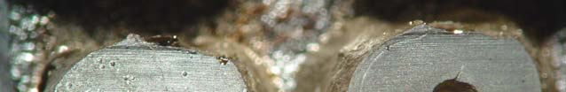
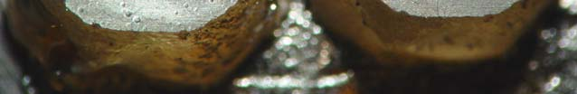
17 Αποτελέσματα Τα αποτελέσματα της πειραματικής μελέτης αναφέρονται συνοπτικά στους πίνακες 1 και 2. Από τα 45 δόντια, παρατηρήθηκαν 11 μικροκενά σε 8 δόντια μεταξύ γουταπέρκας φυράματος οδοντίνης μετά την εκτομή του ακρορριζίου. Η αξιολόγηση έγινε με απλή παρατήρηση σε μεγέθυνση x100 χωρίς καμία ιδιαίτερη ταξινόμηση ως προς το μέγεθος ή την εντόπιση τους. (Εικόνες 1α -1β ) Ρωγμές Ομάδα Α: Σε σύνολο 15 δοντιών παρατηρήθηκαν 7 ρωγμές σε 4 δόντια: μία ολικού μήκους ρωγμή, πέντε ατελείς και μία ρωγμή στην οδοντίνη. Από τις πέντε ατελείς ρωγμές, οι δύο αρχίζουν από τον ριζικό σωλήνα και καταλήγουν στην οδοντίνη και οι υπόλοιπες τρεις αρχίζουν από την εξωτερική επιφάνεια της ρίζας και καταλήγουν στην οδοντίνη. Μετά την εκτομή του ακρορριζίου και πριν την προπαρασκευή της ανάστροφης κοιλότητας παρατηρήθηκαν τρεις ατελείς ρωγμές και μία στην οδοντίνη που μεταβλήθηκαν σε μέγεθος μετά την προπαρασκευή, ενώ μία ολικού μήκους και δύο ατελείς δημιουργήθηκαν μετά την προπαρασκευή. (Εικόνες 2α-2β, 3α-3β, 4α-4β, 5α-5β ) Ομάδα Β: Σε σύνολο 15 δοντιών παρατηρήθηκαν 11 ρωγμές σε 6 δόντια: εννέα ατελείς ρωγμές και δύο ρωγμές στην οδοντίνη. Από τις εννέα ατελείς ρωγμές, οι τέσσερις αρχίζουν από τον ριζικό σωλήνα και καταλήγουν στην οδοντίνη και οι υπόλοιπες πέντε αρχίζουν από την εξωτερική επιφάνεια της ρίζας και καταλήγουν στην οδοντίνη. Μετά την εκτομή του ακρορριζίου και πριν την προπαρασκευή της ανάστροφης κοιλότητας παρατηρήθηκαν έξι ατελείς ρωγμές και μία στην οδοντίνη που μεταβλήθηκαν σε μέγεθος μετά την προπαρασκευή, ενώ τρεις ατελείς και μία στην οδοντίνη δημιουργήθηκαν μετά την προπαρασκευή. (Εικόνες 6α-6β, 7α-7β, 8α-8β) Ομάδα Γ: Ένα δείγμα καταστράφηκε κατά την διάρκεια της επιμετάλλωσης και απομακρύνθηκε. Σε σύνολο 14 δοντιών παρατηρήθηκαν 10 ρωγμές σε 7 δόντια: δύο ολικού μήκους ρωγμές, πέντε ατελείς και τρεις ρωγμές στην οδοντίνη. Από τις πέντε ατελείς ρωγμές, οι τέσσερις αρχίζουν από τον 16
18 ριζικό σωλήνα και καταλήγουν στην οδοντίνη και μία αρχίζει από την εξωτερική επιφάνεια της ρίζας και καταλήγει στην οδοντίνη. Μετά την εκτομή του ακρορριζίου και πριν την προπαρασκευή της ανάστροφης κοιλότητας παρατηρήθηκαν μία ολικού μήκους ρωγμή, δύο ατελείς και δυο στην οδοντίνη που μεταβλήθηκαν σε μέγεθος μετά την προπαρασκευή, ενώ μία ολικού μήκους ρωγμή, τρεις ατελείς και μία στην οδοντίνη δημιουργήθηκαν μετά την προπαρασκευή. Εικόνες 9α-9β-9γ-9δ, 10α-10β, 11α-11β ) Μικροανωμαλίες στα όρια της κοιλότητας Ομάδα Α: Από τα 15 δόντια, εννέα δόντια δεν εμφάνισαν μικροανωμαλίες, τέσσερα εμφάνισαν οριακές μικροανωμαλίες και δύο εμφάνισαν εκτεταμένες μικροανωμαλίες. (Εικόνες 12, 13, 14) Ομάδα Β: Από τα 15 δόντια, τέσσερα δόντια δεν εμφάνισαν μικροανωμαλίες, πέντε εμφάνισαν εντύπωμα, πέντε εμφάνισαν οριακές μικροανωμαλίες, ενώ ένα εμφάνισε εκτεταμένες μικροανωμαλίες. (Εικόνες 15α- 15β, 16α-16β, 17α-17β) Ομάδα Γ: Από τα 14 δόντια, τέσσερα δόντια δεν εμφάνισαν μικροανωμαλίες, ένα εμφάνισε εντύπωμα, πέντε εμφάνισαν οριακές μικροανωμαλίες, ενώ τέσσερα εμφάνισαν εκτεταμένες μικροανωμαλίες. (Εικόνες 18, 19, 20) Στατιστική ανάλυση Από την στατιστική ανάλυση που ακολουθήθηκε δεν βρέθηκαν στατιστικά σημαντικές διαφορές ανάμεσα στην κλασική τεχνική και στις τεχνικές με ρύγχη υπερήχων, όταν αξιολογήθηκαν οι ρωγμές και οι μικροανωμαλίες στην επιφάνεια της οδοντίνης (P >0.05). 17
19 Συζήτηση Η πειραματική μελέτη έγινε σε πρόσθια μονόριζα δόντια, άνω και κάτω γνάθου. Η επιλογή τους έγινε με βάση την μειωμένη πολυπλοκότητα της ανατομίας των ριζικών σωλήνων, την ευθεία πορεία και την ύπαρξη ενός ριζικού σωλήνα κριτήρια που διευκόλυναν την καλύτερη τυποποίηση των δειγμάτων. Τα δόντια που χρησιμοποιήθηκαν ήταν προσφάτως εξαγχθέντα και ιδιαίτερη προσοχή δόθηκε στην συνεχή διατήρηση τους σε υγρό περιβάλλον, με σκοπό να αποφευχθεί η αφυδάτωση της οδοντίνης. Κάτι τέτοιο είναι πιθανό να επηρεάσει τις μηχανικές ιδιότητες της οδοντίνης, με συνέπεια να αυξηθεί το ποσοστό δημιουργίας ρωγμών σε σύγκριση με την οδοντίνη που δεν έχει υποστεί αφυδάτωση. (Kahler 2003) Η συντηρητική ενδοδοντική θεραπεία των δοντιών έγινε πριν την εκτομή και προπαρασκευή των ανάστροφων κοιλοτήτων. Ο Beling et al. (1997), αξιολογώντας τον αριθμό και τον τύπο των ρωγμών πριν και μετά την προπαρασκευή της ανάστροφης κοιλότητας δεν βρήκαν στατιστικά σημαντικές διαφορές ανάμεσα σε δόντια με και χωρίς ενδοδοντική θεραπεία. Αντίθετα, οι Morgan & Marshall (1999) και ο Gondim et al (2002,2003) θεώρησαν ως επιβαρυντικό παράγοντα στην δημιουργία ρωγμών, την αφυδάτωση και την αλλαγή του σχήματος του ριζικού σωλήνα, που υπόκεινται τα δόντια κατά την συντηρητική ενδοδοντική θεραπεία. Με βάση τα παραπάνω και προς αποφυγή τυχόν ψευδών αποτελεσμάτων (artifacts), στην παρούσα μελέτη, τα δόντια εξετάστηκαν προσεκτικά πριν και μετά την έμφραξη του ριζικού σωλήνα μέσω οπτικού μικροσκοπίου και στερεομικροσκοπίου (μεγεθύνσεις x5, x12). Χρησιμοποιήθηκε η τεχνική αντιγραφής των δοντιών με αποτύπωση των επιφανειών με σιλικόνη και πλήρωση με ρητίνη. Η τεχνική αντιγραφής με αρχική λήψη αποτυπωμάτων επιτρέπει την ταυτόχρονη εξέταση και σύγκριση των δειγμάτων του ίδιου δοντιού πριν και μετά την προπαρασκευή της ανάστροφης κοιλότητας, χωρίς καμία καταστροφή του (Crang 1988). Η άμεση τοποθέτηση και εξέταση στο ηλεκτρονικό μικροσκόπιο σάρωσης καταλήγει σε καταστροφή του δοντιού. Επίσης, η αφυδάτωση που υπόκειται το δόντι κατά την άμεση τοποθέτηση του στο SEM σχετίζεται με την δημιουργία ψευδών αποτελεσμάτων (artifacts), με συνέπεια την παρερμηνεία των ρωγμών στην επιφάνεια της ρίζας. (Crang 1988, Torabinejad 1995) Αντίθετοι στην άποψη αυτή έχουν υπάρξει 18
20 ερευνητές, οι οποίοι υποστηρίζουν ότι τα ψευδή αποτελέσματα μπορεί να θεωρηθούν αμελητέας σημασίας. (Janda 1995, Taschieri 2004) Η τεχνική αντιγραφής με πλήρωση του αποτυπώματος λεπτόρρευστης σιλικόνης με εποξυλική ρητίνη μπορεί να θεωρηθεί αρκετά ακριβής και αξιόπιστη. (Walsh 1991) Το ηλεκτρονικό μικροσκόπιο σάρωσης (SEM) χρησιμοποιήθηκε για την παρατήρηση των επιφανειών των αντιγράφων των ακρορριζίων πριν και μετά την προπαρασκευή της ανάστροφης κοιλότητας, δίνοντας δυνατότητα λεπτομερούς απεικόνισης. (Wright 2004) Εκτός του SEM έχουν χρησιμοποιηθεί και άλλα μέσα αξιολόγησης, όπως το απλό μικροσκόπιο, το στερεομικροσκόπιο, το ενδοσκόπιο, με ή χωρίς την προσθήκη χρωστικών και φωτός, τα οποία δεν μπορούν να απεικονίσουν τα δείγματα σε μεγάλες μεγεθύνσεις. Μικρές ατελείς ρωγμές, ρωγμές στην οδοντίνη και μικροανωμαλίες στα όρια της κοιλότητας χρειάζονται μεγεθύνσεις πάνω από x100 φορές για να παρατηρηθούν, πλεονέκτημα που έχει η χρήση του SEM. Σε μικρότερες μεγεθύνσεις, ορισμένες ρωγμές είναι μη ανιχνεύσιμες με αποτέλεσμα να μην συμπεριληφθούν στην αξιολόγηση. (Morgan 1999, Gondim 2002, von Arx 2003) Στην παρούσα μελέτη, έγινε επιμέρους έλεγχος των δειγμάτων σε στερεομικροσκόπιο (μεγέθυνση x12-x32) πριν την τοποθέτηση τους στο SEM, με σκοπό την αρχική παρατήρηση τους και ανίχνευση εμφανών ρωγμών στην επιφάνεια τους. Η εκτομή του ακρορριζικού τμήματος της ρίζας πραγματοποιήθηκε με δίσκο κοπής από διαμάντι προσαρμοσμένο σε μικροτόμο σκληρών οδοντικών ιστών, με γωνία εκτομής 0º μοίρες. Οι επιφάνειες των ριζών βρέθηκαν λείες, με παράλληλες γραμμές και χωρίς έντονες ανωμαλίες, χαρακτηριστικά που διευκόλυναν την παρατήρηση στο ηλεκτρονικό μικροσκόπιο σάρωσης. Η επιφάνεια γουταπέρκας - φυράματος - οδοντίνης ύστερα από παρατήρηση σε μεγέθυνση x100 βρέθηκε λεία, ομαλή, χωρίς ιδιαίτερα κενά ανάμεσα στο εμφρακτικό υλικό και τα τοιχώματα του ριζικού σωλήνα. Επιπρόσθετα, δεν παρατηρήθηκε «επάλειψη» των τοιχωμάτων της οδοντίνης με γουταπέρκα πάνω στην επιφάνεια της ρίζας. Τα παραπάνω ευρήματα συμφωνούν με τα ευρήματα των Weston et al. (1999), οι οποίοι σύγκριναν διάφορους τύπους εγγλυφίδων με δίσκο κοπής προσαρμοσμένο σε μικροτόμο για εκτομή του ακρορριζίου. Η καλύτερη απεικόνιση της επιφάνειας της ρίζας και η αμετάβλητη πρόσφυση των 19
21 εμφρακτικών υλικών με τα τοιχώματα της οδοντίνης, χωρίς την δημιουργία μικροκενών ή την «επάλειψη» της οδοντίνης με γουταπέρκα ήταν χαρακτηριστικά που παρατηρήθηκαν μόνο ύστερα από την εκτομή του ακρορριζίου με δίσκο κοπής. (Weston 1999) Στην παρούσα μελέτη, παρατηρήθηκαν στο 25% των δειγμάτων μικροκενά ανάμεσα στο εμφρακτικό υλικό και τα τοιχώματα της οδοντίνης, ενώ πρέπει να σημειωθεί ότι παρατηρήθηκε σε όλα τα δείγματα μικρή ολίσθηση των εμφρακτικών υλικών κατά την φορά της εκτομής (Εικόνα 1α-1β). Το εύρημα αυτό είναι πιθανό να οφείλεται στην μη τρισδιάστατη ερμητική έμφραξη των ριζικών σωλήνων από την στιγμή που δεν παρατηρήθηκαν άλλες μεταβολές στην μάζα της γουταπέρκας και του φυράματος. Στην κλινική πράξη η χρήση εγγλυφίδων προσαρμοσμένων σε χειρολαβή υψηλών ταχυτήτων για την εκτομή του ακρορριζίου μπορεί να προκαλέσει μεταβολές στην μάζα των εμφρακτικών υλικών, με συνέπεια την δημιουργία νέων μικροκενών ή την αλλαγή του μεγέθους των προϋπαρχόντων. (Nedderman 1988, Gutmann 1993, Gondim 2005) Επίσης, η εκτομή του ακρορριζίου με οποιοδήποτε μέσο μπορεί να αποκαλύψει μικροκενά που έγιναν κατά την έμφραξη του ριζικού σωλήνα. (Kontakiotis 2004, Wu 2001) Τα μικροκενά αποτελούν δίοδο για την έξοδο μικροοργανισμών στους περιακρορριζικούς ιστούς ή εστίες ανάπτυξης μικροβίων. Γι αυτό το λόγο, η προπαρασκευή κοιλότητας για ανάστροφη έμφραξη στο ακρορριζικό τριτημόριο του ριζικού σωλήνα κρίνεται αναγκαία. (Kim 2001, 2006) Οι τεχνικές προπαρασκευής κοιλότητας για ανάστροφη έμφραξη είναι ένα στάδιο της χειρουργικής ενδοδοντίας που έχει απασχολήσει ιδιαίτερα την επιστημονική κοινότητα τα τελευταία χρόνια, ύστερα από την εισαγωγή των υπερήχων. H κλασική τεχνική παρουσιάζει πολλά μειονεκτήματα για τον επεμβαίνοντα, τα οποία αναφορικά είναι: α. η γωνία εκτομής του ακρορριζίου γίνεται πολύ μεγάλη (45º-60º) για ευκολότερη προσπέλαση, με αποτέλεσμα μεγάλη έκθεση οδοντινικών σωληναρίων και πιθανή αποτυχία απομάκρυνσης ανατομικών δομών, όπως παράπλευροι ριζικοί σωλήνες και ακρορριζικά δέλτα, β. η προσπέλαση στην περιοχή του ακρορριζίου είναι δύσκολη, ειδικότερα όταν το οπτικό πεδίο είναι περιορισμένο, με συνέπεια μεγάλη αποκοπή οστίτη ιστού, γ. η προπαρασκευή της κοιλότητας δεν ακολουθεί τον επιμήκη άξονα της ρίζας, με αποτέλεσμα να αυξάνονται οι πιθανότητες διάτρησης και να μειώνεται η συγκράτηση των εμφρακτικών υλικών της ανάστροφης κοιλότητας, δ. η προπαρασκευή προκαλεί υπέρμετρη διεύρυνση της κοιλότητας, με αποτέλεσμα 20
22 την εξασθένιση την ακρορριζικής οδοντίνης ε. ο νεκρωτικός ιστός που βρίσκεται στους ισθμούς δεν μπορεί να απομακρυνθεί. (Carr 1997, Kim 1997, 2001) Η εισαγωγή των υπερήχων για την προπαρασκευή ανάστροφης κοιλότητας έγινε για να αντιμετωπιστούν όλα τα παραπάνω προβλήματα που δημιουργούνται με την κλασική τεχνική. Περιληπτικά τα πλεονεκτήματα των υπερήχων έναντι της κλασικής τεχνικής είναι: α. ευκολότερη και καλύτερη προσπέλαση, κυρίως σε περιοχές με περιορισμένο οπτικό πεδίο, β. πιο συντηρητική προπαρασκευή της ανάστροφης κοιλότητας, η οποία ακολουθεί τον επιμήκη άξονα της ρίζας, γ. καλύτερη απομάκρυνση οδοντικού τρίματος (debris) από τα τοιχώματα της κοιλότητας, δ. ακριβείς προπαρασκευές με παράλληλα τοιχώματα και καλύτερη συγκράτηση των εμφρακτικών υλικών, ακόμα και σε ακρορριζικούς ισθμούς. (Kim 2001) Σε αντίθεση με τα παραπάνω πλεονεκτήματα οι υπέρηχοι έχουν ενοχοποιηθεί για την δημιουργία μεγαλύτερου αριθμού ρωγμών στην επιφάνεια του ακρορριζίου και μικροανωμαλιών στα όρια της ανάστροφης κοιλότητας, σε σύγκριση με την κλασική τεχνική. Η δημιουργία ρωγμών σχετίζεται με την πιθανότητα κατάγματος της ρίζας σε δεύτερο χρόνο, την ανεπαρκή έμφραξη της ανάστροφης κοιλότητας και την πιθανότητα μικροβιακής επιμόλυνσης, ενώ η δημιουργία μικροανωμαλιών στα όρια της κοιλότητας σχετίζεται με την ικανότητα πρόσφυσης των εμφρακτικών υλικών και την μικροδιείσδυση στις περιοχές αυτές. (Gray 2000, Gondim 2003) Ένας αριθμός ερευνών σε εξαγχθέντα δόντια έχει δείξει μεγαλύτερο ποσοστό δημιουργίας ρωγμών μετά την προπαρασκευή ανάστροφης κοιλότητας με ρύγχη υπερήχων, σε σύγκριση με εγγλυφίδες προσαρμοσμένες σε μικροχειρολαβή. (Saunders 1994, Abedi 1995, Frank 1996, Khabbaz 2004) Oι Saunders et al.(1994) παρατήρησαν εξαγχθέντα δόντια σε SEΜ ύστερα από αφυδάτωση τους και αξιολόγησαν την τεχνική υπερήχων με ρύγχη ανοξείδωτου χάλυβα χωρίς διαμάντι στο κοπτικό άκρο, αναφέροντας μεγαλύτερο ποσοστό ρωγμών στην επιφάνεια του ακρορριζίου σε σχέση με την κλασική τεχνική. Μικρότερο ποσοστό ρωγμών ανέφερε ο Abedi et al. (1995), ο οποίος χρησιμοποιώντας την τεχνική αντιγραφής σε εξαγχθέντα δόντια μετά από παρατήρηση σε SEM (μεγέθυνση x30), σύγκρινε τις δύο τεχνικές και ανέφερε αυξημένο ποσοστό ρωγμών με την τεχνική υπερήχων με ρύγχη ανοξείδωτου 21
23 χάλυβα χωρίς διαμάντι στο κοπτικό άκρο, ειδικότερα σε περιοχές όπου η οδοντίνη που περιβάλλει την κοιλότητα είναι πολύ λεπτή (< 2mm). Παρόμοια ευρήματα ανέφεραν ο Frank et al. (1996) παρατηρώντας όμως τις επιφάνειες των δοντιών με οπτικό μικροσκόπιο σε μεγέθυνση μέχρι x16 φορές, ύστερα από χρώση με κυανό του μεθυλενίου. Ο Min et al. (1999) σύγκριναν την κλασική τεχνική με την τεχνική υπερήχων με ρύγχη ανοξείδωτου χάλυβα με ή χωρίς διαμάντι στο κοπτικό άκρο, παρατηρώντας τις επιφάνειες με μικροσκόπιο confocal (x40,) και στην συνέχεια παίρνοντας τομές και κάνοντας ιστολογική εξέταση με εκ νέου παρατήρηση σε confocal μικροσκόπιο (x40, x100). Bρήκαν μεγαλύτερο αριθμό ρωγμών μετά την χρήση ρυγχών υπερήχων χωρίς διαμάντι στο κοπτικό άκρο, έναντι των άλλων τεχνικών. (Min 1999) Ο Κhabbaz et al. (2004) σύγκριναν την κλασική τεχνική με την τεχνική υπερήχων με ρύγχη ανοξείδωτου χάλυβα με ή χωρίς διαμάντι στο κοπτικό άκρο και ρύγχος σε ηχητική συχνότητα (Sonic retro-prep tip) και βρήκαν μεγαλύτερο αριθμό ρωγμών μετά την χρήση ρυγχών υπερήχων χωρίς διαμάντι στο κοπτικό άκρο, έναντι των άλλων τεχνικών. Συγκριτικά με τις προηγούμενες έρευνες ο συνολικός αριθμός ρωγμών ήταν αρκετά μικρότερος σε όλες τις τεχνικές. Η παρατήρηση έγινε μέσω ψηφιακής απεικόνισης (ψηφιακή μεγέθυνση x100), ύστερα από φωτογράφηση των επιφανειών με ψηφιακή κάμερα. (Κhabbaz 2004) Οι παραπάνω έρευνες συμφωνούν ότι η χρήση της τεχνικής υπερήχων με ρύγχος ανοξείδωτου χάλυβα χωρίς διαμάντι στο κοπτικό άκρο προκαλεί μεγαλύτερο αριθμό ρωγμών σε σχέση με την τεχνική υπερήχων με ρύγχος ανοξείδωτου χάλυβα με διαμάντι στο κοπτικό άκρο και με την κλασική τεχνική. Αντίθετα, άλλες έρευνες που συγκρίνουν την κλασική τεχνική με αυτές των υπερήχων, δεν αναφέρουν αξιοσημείωτες διαφορές στον αριθμό ρωγμών. Ο Lloyd et al. (1996), χρησιμοποιώντας την τεχνική αντιγραφής σε εξαγχθέντα δόντια μετά από παρατήρηση σε SEM (μεγέθυνση x20, x80), δεν βρήκε στατιστικά σημαντικές διαφορές ανάμεσα σε κοιλότητες που προπαρασκευάστηκαν με την κλασική τεχνική και σε κοιλότητες που προπαρασκευάστηκαν με ρύγχος σε ηχητικές συχνότητες (Sonic Retro-prep tip). Ο Rainwater et al. (2000) σύγκριναν την κλασική τεχνική με την τεχνική υπερήχων με ρύγχη με ή χωρίς διαμάντι στο κοπτικό άκρο, ύστερα από 22
24 παρατήρηση σε οπτικό μικροσκόπιο σε μεγέθυνση x20, χωρίς να βρουν στατιστικά σημαντικές διαφορές. (Lloyd et al.1996, Rainwater et al. 2000) Απουσία ρωγμών έχει αναφερθεί σε δύο έρευνες. (Waplington 1997, Lin 1999) O Waplington et al. (1997) σύγκριναν την κλασική τεχνική και την τεχνική υπερήχων με ρύγχη χωρίς διαμάντι στο κοπτικό άκρο, χρησιμοποιώντας την τεχνική αντιγραφής σε εξαγχθέντα δόντια μετά από παρατήρηση σε SEM. Ο Lin et al. (1999) σύγκριναν τις ίδιες τεχνικές, χρησιμοποιώντας στερεομικροσκόπιο σε μεγέθυνση x30, αφού είχε προηγηθεί χρώση των επιφανειών με κυανό του μεθυλενίου και χωρίς να έχει γίνει συντηρητική ενδοδοντική θεραπεία πριν την προπαρασκευή των ανάστροφων κοιλοτήτων. Έρευνες έχουν συγκρίνει και διάφορα είδη ρυγχών υπέρηχων μεταξύ τους, σε ίδιες ή διαφορετικές συχνότητες, σε δόντια με προπαρασκευασμένους ή μη ριζικούς σωλήνες, στην προσπάθεια τους να αξιολογήσουν την παρουσία ρωγμών στην επιφάνεια του ακρορριζίου. (Zuolo 1999, Layton 1996, Navarre 2002, Brent 1999, Gondim 2002, Beling 1997, Rainwater 2000, Ishikawa 2003, Taschieri 2004) Οι Layton et al. (1996) και Beling et al. (1997) στις μελέτες τους χρησιμοποίησαν το ίδιο ερευνητικό μοντέλο, αλλά βρήκαν διαφορετικά αποτελέσματα. Οι Layton et al.(1996), παρατηρώντας δόντια με μη προπαρασκευασμένους ριζικούς σωλήνες μετά από την προπαρασκευή ανάστροφης κοιλότητας με ρύγχος υπερήχων χωρίς διαμάντι στο κοπτικό άκρο, σε χαμηλή ισχύ, βρήκε αυξημένο αριθμό ρωγμών (30%) στην επιφάνεια των ακρορριζίων. Από την άλλη πλευρά ο Beling et al.(1997), σε παρόμοιες συνθήκες, βρήκε πολύ μικρότερο ποσοστό (10%). Τα αποτελέσματα των δύο ομάδων ερευνητών συμφωνούν όταν χρησιμοποιήθηκε υψηλή ισχύς υπερήχων, όπου βρέθηκε συνολικά χαμηλό ποσοστό ρωγμών. Στατιστικά σημαντικές διαφορές δεν βρέθηκαν σε μελέτες που σύγκριναν ρύγχη υπερήχων με και χωρίς διαμάντι στο κοπτικό άκρο σε ίδια ισχύ. (Zuolo 1999, Navarre 2002, Brent 1999, Rainwater 2000) Επίσης, σε άλλες μελέτες συγκρίθηκαν οι τεχνικές προπαρασκευής ανάστροφης κοιλότητας με την χρήση ρυγχών με ή χωρίς διαμάντι και ρυγχών με νιτρικό ζιρκόνιο στο κοπτικό άκρο και δεν βρέθηκαν στατιστικά σημαντικές διαφορές στο ποσοστό ρωγμών στην επιφάνεια του ακρορριζίου, όταν η ισχύς ρυθμίστηκε στην ίδια τιμή για όλες τις τεχνικές. (Gondim 2002, Beling 1997, Rainwater 2000, Taschieri 2004) 23
25 Με βάση τα αποτελέσματα των προηγούμενων ερευνών γίνεται αντιληπτό ότι η ρύθμιση της ισχύος του υπερήχου σε υψηλή τιμή μπορεί να καταλήξει σε αύξηση του αριθμού των ρωγμών (Frank 1995, Taschieri 2004), κάτι που μπορεί να παρατηρηθεί και σε χαμηλή ισχύ. (Layton 1996, Ishikawa 2003) Τα αποτελέσματα αυτά μπορούν να δικαιολογηθούν από το γεγονός ότι οι συχνότητες που λειτουργούν οι υπέρηχοι (30-40 kηz) προκαλούν μεγάλου πλάτους δόνηση, είτε χρησιμοποιηθούν σε χαμηλή ή σε υψηλή ισχύ, με συνέπεια την πρόκληση εγκάρσιας ταλάντωσης στην περιοχή του ακρορριζίου. Η ταλάντωση αυτή μαζί με την ενέργεια που παράγεται κατά την επαφή του ρύγχους υπερήχων με τα τοιχώματα της οδοντίνης μπορεί να αυξήσει το ποσοστό ρωγμών στη ρίζα. (Lin 1999, Walmsley 1996) Οι περιοδοντικοί ιστοί σε in vivo συνθήκες μπορούν να απορροφήσουν μέρος της συνολικής ενέργειας που δημιουργείται κατά την χρήση των υπερήχων και να μειωθεί ο αριθμός των ρωγμών. (Min 1997, Calzonetti 1998) Ο Min et al.(1997), θεωρώντας σημαντική αυτήν την παράμετρο, πρότεινε την χρήση ανθρώπινων πτωμάτων (in situ) για καλύτερη προσομοίωση με τις in vivo συνθήκες, λαμβάνοντας πάντα υπ όψιν το γεγονός ότι οι περιοδοντικοί ιστοί έχουν υποστεί επιμέρους αφυδάτωση και δεν ομοιάζουν πλήρως με τους in vivo περιοδοντικούς ιστούς. Σε έρευνα που έγινε σε ανθρώπινα πτώματα και αξιολογήθηκε η παρουσία ρωγμών μετά από την προπαρασκευή ανάστροφων κοιλοτήτων με δύο είδη ρυγχών ανοξείδωτου χάλυβα χωρίς διαμάντι στο κοπτικό άκρο, δεν βρέθηκε καμία ρωγμή στα δείγματα. Η παρατήρηση έγινε σε SEM (x25,50,100) ύστερα από αποτύπωση των ακρορριζίων με σιλικόνη και χωρίς να ακολουθηθεί η τεχνική αντιγραφής με ρητίνη. (Calzonetti 1998) Παρόμοια αποτελέσματα βρήκε ο Gray et al. (2000) συγκρίνοντας την κλασική τεχνική με την τεχνική υπερήχων με ρύγχος ανοξείδωτου χάλυβα χωρίς διαμάντι στο κοπτικό άκρο, σε χαμηλή και υψηλή ισχύ και σε δόντια χωρίς ενδοδοντική θεραπεία. Το ποσοστό ρωγμών θεωρήθηκε αμελητέο, ενώ η παρατήρηση των ακρορριζίων έγινε σε SEM (x30) ύστερα από την εφαρμογή της τεχνικής αντιγράφων. Οι De Bryne & De Moor (2005) σε μια νεότερη μελέτη σε ανθρώπινα πτώματα σύγκριναν ρύγχη υπερήχων ανοξείδωτου χάλυβα με διαμάντι κοπής στο κοπτικό άκρο σε μέση και χαμηλή ισχύ, σε προπαρασκευές ανάστροφων κοιλοτήτων σε εξαγχθέντα δόντια και δόντια πτωμάτων. Κατέληξαν στο 24
26 συμπέρασμα ότι η χρήση ρυγχών υπερήχων σε μέση ισχύ προκαλεί αμελητέο αριθμό ρωγμών σε δόντια πτωμάτων έναντι των εξαγχθέντων, ενώ η χρήση ρυγχών υπερήχων σε χαμηλή ισχύ δεν προτείνεται καθώς προκάλεσε αυξημένο αριθμό ρωγμών και στις δύο κατηγορίες. Η παρατήρηση έγινε σε SEM (x20,35) ύστερα από αποτύπωση των ακρορριζίων με σιλικόνη και χωρίς να ακολουθηθεί η τεχνική αντιγραφής με ρητίνη. (De Bryne & De Moor 2005) Στην παρούσα μελέτη χρησιμοποιήθηκε μέση ισχύς με βάση τις οδηγίες του κατασκευαστή και λαμβάνοντας υπ όψιν τα αποτελέσματα των προηγουμένων ερευνών, καθώς και τα αποτελέσματα άλλων ερευνητών που σύγκριναν υψηλή ή χαμηλή ισχύ υπερήχων, έναντι της μέσης ισχύος και βρήκαν μικρότερο ποσοστό ρωγμών όταν χρησιμοποιήθηκε η μέση ισχύς. (De Bryne & De Moor 2005, Gondim 2002) Στην παρούσα μελέτη έγινε σύγκριση της κλασικής τεχνικής με εγγλυφίδα Νο1 προσαρμοσμένη σε μικροχειρολαβή χαμηλών ταχυτήτων και της τεχνικής υπερήχων με ρύγχος ανοξείδωτου χάλυβα με και χωρίς διαμάντι στο κοπτικό άκρο. Δεν βρέθηκε στατιστικά σημαντική διαφορά στον αριθμό των ρωγμών στην επιφάνεια των ακρορριζίων ανάμεσα στις τρεις τεχνικές, παρά το ότι οι τεχνικές των υπερήχων προκάλεσαν περισσότερες ρωγμές στο σύνολο έναντι της κλασικής τεχνικής. Σε ποσοστό 60% των δειγμάτων εμφανίστηκαν ρωγμές, όμως το ποσοστό των ρωγμών ανά δείγμα μειώνεται στο 27% με την κλασική τεχνική, 40% με ρύγχος υπερήχων με διαμάντι στο κοπτικό άκρο και 50% με ρύγχος υπερήχων χωρίς διαμάντι στο κοπτικό άκρο. Το ποσοστό νέων ρωγμών μετά την προπαρασκευή ανάστροφων κοιλοτήτων βρέθηκε, 20%, 27%, 33% για τις τρεις τεχνικές, αντίστοιχα. Επίσης, έγινε ταξινόμηση των ρωγμών με σκοπό τον καλύτερο προσδιορισμό και αξιολόγηση τους και την συσχέτιση τους με κάθε τεχνική. Η μεγαλύτερη διαφορά παρουσιάστηκε στο σύνολο των ατελών ρωγμών, σε ποσοστό 42%, έναντι 7% των ολικού μήκους ρωγμών και 13% των ρωγμών στην οδοντίνη, ένα εύρημα που μπορεί να δικαιολογηθεί από την άμεση γειτνίαση και επαφή του ρύγχους με τα τοιχώματα της οδοντίνης κατά την προπαρασκευή της κοιλότητας. Αξιοσημείωτο εύρημα μπορεί να θεωρηθεί η απουσία νέας ατελούς ρωγμής που αρχίζει από την εξωτερική επιφάνεια της ρίζας και καταλήγει στην οδοντίνη ( ατελής τύπου β ), μετά την προπαρασκευή ανάστροφης κοιλότητας με οποιαδήποτε τεχνική. Οι ατελείς ρωγμές τύπου β που παρατηρήθηκαν μετά την 25
27 εκτομή του ακρορριζίου και πριν την προπαρασκευή της ανάστροφης κοιλότητας είναι πιθανό να προκλήθηκαν είτε κατά την βίαιη εξαγωγή των δοντιών ή κατά την εκτομή του ακρορριζίου. (Rainwater 2000) Στην ομάδα Α από τις 7 συνολικά ρωγμές που βρέθηκαν, οι 4 εντοπίστηκαν στη στενότερη περιοχή του ακρορριζίου, στην ομάδα Β από τις 11 ρωγμές εντοπίστηκαν οι 5 και από την ομάδα Γ από τις 10 ρωγμές εντοπίστηκαν οι 3, αντίστοιχα. Τα αποτελέσματα δείχνουν ότι μόνο ένας μικρός συνολικά αριθμός ρωγμών εντοπίζεται στις στενές περιοχές του ακρορριζίου και δεν επιβεβαιώνει τον κίνδυνο πρόκλησης περισσοτέρων ρωγμών σε μικρής διαμέτρου ακρορρίζια, κάτι που έχει υποστηριχτεί από προηγούμενους ερευνητές. (Abedi 1995, Frank 1996) Αντίθετα, τα αποτελέσματα αυτά συμφωνούν με άλλους ερευνητές που καταλήγουν στο συμπέρασμα ότι η δημιουργία ρωγμών δεν σχετίζεται με μικρές διαστάσεις του ακρορριζίου. (De Bryne & De Moor 2005, Gondim 2002) Επιπρόσθετα, δεν μπορεί να υπάρξει συσχέτιση μεταξύ του τύπου των ρωγμών και του είδους της τεχνικής που ακολουθήθηκε, καθώς δεν παρατηρήθηκε σημαντικού βαθμού συσχέτιση ανάμεσα στην πρόκληση συγκεκριμένου τύπου ρωγμής και τεχνικής. Τέλος, πρέπει να σημειωθεί ότι η ταξινόμηση των ρωγμών έγινε σε σχέση με την εντόπιση στις επιφάνειες των ακρορριζίων χωρίς να αξιολογηθεί το βάθος των ρωγμών μέσα στην οδοντίνη, καθώς υπήρξε δυσκολία στην αντικειμενική μέτρηση αυτής της διάστασης με το SEM. Η σημασία των μικροανωμαλιών στα όρια των ανάστροφων κοιλοτήτων έχει συσχετιστεί με την πρόσφυση των εμφρακτικών υλικών στις επιφάνειες των τοιχωμάτων της οδοντίνης και την πιθανή μικροδιείσδυση μικροοργανισμών. Ένας αριθμός ιn vitro ερευνών καταλήγει στο συμπέρασμα ότι η παρουσία μικροανωμαλιών στα όρια των ανάστροφων κοιλοτήτων δεν επηρεάζει αρνητικά την πρόσφυση των εμφρακτικών υλικών, χωρίς να προκαλεί αύξηση της ακρορριζικής μικροδιείσδυσης. 13,33,34 Οι μικροανωμαλίες στα όρια μετά από την προπαρασκευή ανάστροφης κοιλότητας έχουν ερευνηθεί σε διάφορες πειραματικές μελέτες. (Layton 1996, Lloyd 1996,, Wapligton 1997, Gray 2000, Rainwater 2000, Navarre 2002, Gondim 2002, Ishikawa 2003, Taschieri 2004,De Bryne & De Moor 2005) Οι ερευνητές, σε in vitro μελέτες, καταλήγουν στο ομόφωνο συμπέρασμα ότι η 26
28 χρήση υπερήχων αυξάνει το ποσοστό μικροανωμαλιών στα όρια της ανάστροφης κοιλότητας, όταν συγκρίνεται με την κλασική τεχνική. (Layton 1996, Lloyd 1996, Wapligton 1997) Το ποσοστό μικροανωμαλιών στα όρια της κοιλότητας μετά την χρήση εγγλυφίδας, στην κλινική πράξη, αυξάνεται καθώς η προσπέλαση είναι πολύ πιο δύσκολη σε σχέση με τις in vitro συνθήκες. (Lloyd 1996, Gray 2000, Taschieri 2004)Σε συγκρίσεις που έγιναν μεταξύ διαφορετικών τύπων ρυγχών υπερήχων δεν έχουν παρατηρηθεί στατιστικά σημαντικές διαφορές (Navarre 2002, De Bryne 2005, Taschieri 2004, Rainwater 2000, Ishikawa 2003), ενώ μία έρευνα του Zuolo et al. (1999) βρήκε μικροανωμαλίες στα όρια κοιλοτήτων που προπαρασκευάστηκαν με ρύγχος με διαμάντι στο κοπτικό άκρο σε αντίθεση με εκείνες που προπαρασκευάστηκαν με ρύγχος χωρίς διαμάντι, οι οποίες δεν εμφάνισαν μικροανωμαλίες στα όρια. Ο Gondim et al. (2002) κατέληξαν σε αντίθετα αποτελέσματα, βρίσκοντας μεγαλύτερη συνολική επιφάνεια (mm²) μικροανωμαλιών στα όρια κοιλοτήτων που προπαρασκευάστηκαν με ρύγχη χωρίς διαμάντι στο κοπτικό άκρο. Οι μικροανωμαλίες στα όρια κοιλοτήτων έχουν αξιολογηθεί και σε μελέτες που έγιναν σε ρύγχη υπερήχων σε διαφορετικές τιμές ισχύος. (Frank 1995, Layton 1996, Wapligton 1997, Gray 2000, Taschieri 2004, De Bryne 2005) Το ποσοστό μικροανωμαλίων αυξήθηκε όταν χρησιμοποιήθηκε υψηλή ή χαμηλή ισχύς, ενώ αντίθετα σε έρευνα του Gray et al. (2000) σε ανθρώπινα πτώματα το ποσοστό παρέμεινε στα ίδια επίπεδα όταν αυξήθηκε η ισχύς του υπερήχου. Οι ίδια ομάδα ερευνητών βρήκε μη στατιστικά σημαντική διαφορά στο ποσοστό μικροανωμαλιών ανάμεσα σε κοιλότητες που προπαρασκευάστηκαν με την κλασική τεχνική και σε κοιλότητες που προπαρασκευάστηκαν με την τεχνική υπερήχων, όταν τα δείγματα προέρχονταν από ανθρώπινα πτώματα και πολύ μικρότερο ποσοστό σε κοιλότητες με την κλασική τεχνική έναντι αυτών με την τεχνική υπερήχων, όταν τα δείγματα ήταν εξαγχθέντα δόντια. Η διαφορά στις συνθήκες εφαρμογής των τεχνικών ανάμεσα σε δόντια ανθρώπινων πτωμάτων και σε εξαγχθέντα δόντια είναι μία παράμετρος που δικαιολογεί αυτά τα αποτελέσματα. (Gray 2000) Στην παρούσα μελέτη, παρατηρήθηκε μεγαλύτερο ποσοστό μικροανωμαλιών στα όρια των κοιλοτήτων που προπαρασκευάστηκαν με την τεχνική υπερήχων έναντι της κλασικής τεχνικής. Μεταξύ των ρυγχών υπερήχων δεν παρατηρήθηκαν στατιστικά σημαντικές διαφορές στη δημιουργία 27
29 μικροανωμαλιών στα όρια των ανάστροφων κοιλοτήτων, παρά το γεγονός ότι το μεγαλύτερο σύνολο βαθμών παρατηρήθηκε με την χρήση ρυγχών χωρίς διαμάντι στο κοπτικό άκρο. Η μειωμένη κοπτική ικανότητα του και ο μεγαλύτερος χρόνος προπαρασκευής, που ισοδυναμεί με μεγαλύτερης διάρκειας ταλάντωση και ενέργεια του ρύγχους κατά την επαφή με τα τοιχώματα της οδοντίνης, μπορεί να θεωρηθούν παράγοντες μεγαλύτερου συνόλου βαθμών. Αξιοσημείωτο εύρημα αποτελεί η παρουσία χαρακτηριστικών εντυπωμάτων στα δείγματα των οποίων οι προπαρασκευές έγιναν με ρύγχος υπερήχων με διαμάντι στο κοπτικό άκρο. Η γωνία 90º μοιρών, κοίλου σχήματος, του κοπτικού άκρου με το υπόλοιπο στέλεχος, σε συνδυασμό με την επικάλυψη του ρύγχους με διαμάντι στο σημείο γωνίωσης, είναι στοιχεία που δικαιολογούν την ύπαρξη εντυπωμάτων. Ο Carr (1997)προτείνει την χρήση δύο ρυγχών για την προπαρασκευή ανάστροφης κοιλότητας, με αρχική χρήση του CT-1 και ολοκλήρωση της κοιλότητας σε βάθος και εύρος με το CT-2 ή το CT-3. Τα τελικά ρύγχη CT-2 ή CT-3 έχουν μεγαλύτερες γωνίες και η χρήση τους μπορεί να συμβάλει στην αποφυγή δημιουργίας εντυπωμάτων. Στην μελέτη που έγινε χρησιμοποιήθηκε για πρώτη φορά μόνο το ρύγχος CT-1 με σκοπό την μείωση του χρόνου προπαρασκευής, καθώς διαπιστώθηκε ότι η χρήση ενός μόνο ρύγχους αρκεί για να προπαρασκευαστεί η ανάστροφη κοιλότητα σε βάθος 3mm και να απομακρυνθούν συγχρόνως τα εμφρακτικά υλικά από τα τοιχώματα της κοιλότητας. Η μείωση της πίεσης που ασκείται κατά την χρήση του ρύγχους υπερήχων με φορά προς τον επιμήκη άξονα της ρίζας, συγχρόνως με τον καλύτερο έλεγχο του την χρονική στιγμή που φτάνει σε βάθος 3mm, είναι εφαρμογές οι οποίες παρατηρήθηκε ότι μπορούν να μειώσουν το ποσοστό εντυπωμάτων. Η προπαρασκευή της ανάστροφης κοιλότητας με την κλασική τεχνική ήταν μικρότερης διάρκειας, ενώ η προπαρασκευή με το ρύγχος υπερήχων με διαμάντι στο κοπτικό άκρο υπερτερεί χρονικά σε σχέση με το ρύγχος χωρίς διαμάντι, ευρήματα που συμφωνούν και με προηγούμενες μελέτες. (Sultan 1995, Ishikawa 2003, Taschieri 2004, Khabbaz 2004,) Όλα τα στάδια πραγματοποιήθηκαν από έναν ερευνητή έτσι ώστε να περιοριστεί στο ελάχιστο η μεταβλητή που προκύπτει κατά την παρέμβαση και άλλου επεμβαίνοντα. 28
30 Είναι δύσκολο να συγκριθούν τα αποτελέσματα των ερευνών όταν έχουν ακολουθηθεί ανόμοια πειραματικά μοντέλα. Οι παράμετροι που προκύπτουν σε όλες τις in vitro και in situ μελέτες είναι ανασταλτικοί ως προς την αντικειμενική αξιολόγηση των ρωγμών στην επιφάνεια των ακρορριζίων και των μικροανωμαλιών στα όρια των κοιλοτήτων. Σημαντικοί παράμετροι όπως, η ισχύς του υπερήχου, οι διαφορές σε in vitro και in situ συνθήκες, η μέθοδος παρατήρησης και αξιολόγησης, η προετοιμασία των δειγμάτων, καθώς και επιμέρους παράμετροι όπως, ο χρόνος προπαρασκευής, οι διαφορετικές διαστάσεις των ακρορριζίων, η προπαρασκευή και έμφραξη των ριζικών σωλήνων, οι διαφορές στα είδη των ρυγχών και στις συσκευές υπερήχων, η γωνία εκτομής του ακρορριζίου και η εμπειρία του επεμβαίνοντα, απεικονίζουν την αυξημένη μεταβλητότητα των αποτελεσμάτων. Η ανάγκη τυποποίησης των πειραματικών μοντέλων με ένα ενιαίο πρωτόκολλο θα συμβάλλει στην μείωση του αριθμού ετερογενών ερευνών, με επακόλουθο την σύγχρονη μείωση ψευδών συμπερασμάτων. (Taschieri 2004) Η κλινική συσχέτιση των αποτελεσμάτων δεν έχει ακόμα αποσαφηνιστεί και η επίδραση των ρωγμών και των μικροανωμαλιών στην επιφάνεια του ακρορριζίου, σε σχέση με την επουλωτική ικανότητα των περιακρορριζικών ιστών και την ακρορριζική μικροδιείσδυση χρειάζεται περαιτέρω διερεύνηση. Ελάχιστες κλινικές μελέτες έχουν δημοσιευθεί, συγκρίνοντας την κλασική τεχνική με την τεχνική των υπερήχων αναδρομικά και κατέληξαν σε σημαντικά υψηλότερο ποσοστό επιτυχίας όταν οι προπαρασκευές έγιναν με την τεχνική των υπερήχων, έναντι της κλασικής τεχνικής, διακύμανση που κυμαίνεται στο 17 30%. (Testori 1999, von Arx 2001, Bader 1998)Η εφαρμογή της τεχνικής υπερήχων, συγχρόνως με την προσθήκη νέων υλικών, μικροσκοπίου και μικροχειρουργικών εργαλείων, εξελίσσουν την, μέχρι πρότινος, χειρουργική ενδοδοντία σε μικροχειρουργική ενδοδοντία. (Kim 2006) Τα ποσοστά επιτυχίας φτάνουν στο 96,8% μετά από επανέλεγχο ύστερα από ένα χρόνο και στο 91% μετά από 5-7 χρόνια (Rubinstein 1999, 2002), ενώ άλλες ομάδες ερευνητών ανέφεραν ποσοστό επιτυχίας 95%, 92% και 91% (Taschieri 2006, Chong 2003, Tsesis 2006) Περισσότερες κλινικές μελέτες πρέπει να πραγματοποιηθούν, με σκοπό την ανίχνευση και αξιολόγηση ρωγμών και μικροανωμαλιών σε in vivo συνθήκες, 29
31 την συσχέτιση τους με αποίκιση και μικροδιείσδυση μικροοργανισμών και την σύγκριση τους με in vitro αποτελέσματα. 30
32 Συμπεράσματα 1. Δεν βρέθηκε στατιστικά σημαντική διαφορά στον αριθμό και το είδος των ρωγμών στην επιφάνεια των ακρορριζίων ανάμεσα στις τρεις τεχνικές. 2. Δεν βρέθηκε στατιστικά σημαντική διαφορά στον αριθμό και το είδος μικροανωμαλιών στα όρια των κοιλοτήτων που προπαρασκευάστηκαν με τις τεχνικές υπερήχων έναντι της κλασικής τεχνικής. 3. Μεταξύ των ρυγχών υπερήχων δεν παρατηρήθηκαν στατιστικά σημαντικές διαφορές στη δημιουργία μικροανωμαλιών στα όρια των ανάστροφων κοιλοτήτων. 4. Η εκτομή του ακρορριζίου προκάλεσε ολίσθηση των εμφρακτικών υλικών σε όλα τα δείγματα. Παρατηρήθηκαν μικροκενά ανάμεσα στα εμφρακτικά υλικά και τα τοιχώματα της οδοντίνης σε ορισμένα δείγματα. 31
33 Πίνακες Πίνακας 1. Αποτελέσματα αξιολόγησης ρωγμών Ομάδα Α (εγγλυφίδα Νο1) Ομάδα Β (CT1-D) Ομάδα Γ (CT1) Ολικού μήκους ρωγμές Ατελείς ρωγμές Ατελείς τύπου α Ατελείς τύπου β Ρωγμές στην οδοντίνη Σύνολο Εγγλυφίδα Νο1: Στρογγύλη εγγλυφίδα Νο1 προσαρμοσμένη σε μικροχειρολαβή, CT1-D: ρύγχος υπερήχων ανοξείδωτου χάλυβα με διαμάντι στο κοπτικό άκρο, CT1: ρύγχος υπερήχων ανοξείδωτου χάλυβα χωρίς διαμάντι στο κοπτικό άκρο. Ατελής α: ξεκινάνε από τον ριζικό σωλήνα και καταλήγουν στην οδοντίνη, Ατελής β: ξεκινάνε από την εξωτερική επιφάνεια της ρίζας και καταλήγουν στην οδοντίνη 32
34 Πίνακας 2. Αποτελέσματα της αξιολόγησης των μικροανωμαλιών Ομάδα Α (εγγλυφίδα Νο1) Ομάδα Β (CT1-D) Ομάδα Γ (CT1) Καμία μικροανωμαλία (βαθμοί 0) Εντύπωμα (βαθμός 1) Οριακές μικροανωμαλίες (βαθμοί 2) Εκτεταμένες μικροανωμαλιές (βαθμοί 3) Σύνολο (σύνολο βαθμών) 14* 18* 23* Εγγλυφίδα Νο1: Στρογγύλη εγγλυφίδα Νο1 προσαρμοσμένη σε μικροχειρολαβή, CT1-D: ρύγχος υπερήχων ανοξείδωτου χάλυβα με διαμάντι στο κοπτικό άκρο, CT1: ρύγχος υπερήχων ανοξείδωτου χάλυβα χωρίς διαμάντι στο κοπτικό άκρο. Σύνολο ομάδας Α: (4x2)+(2x3)=14, σύνολο ομάδας Β: (5x1)+(5x2)+(1x3)=18, σύνολο ομάδας Γ: (1x1)+(5x2)+(4x3)= 23 33
35 Βιβλιογραφία Abedi HR, Van Mierlo BL, Wilder-Smith P, Torabinejad M. Effects of ultrasonic root-end cavity preparation on the root apex. Oral Surg Oral Med Oral Pathol Oral Radiol Endod Aug;80(2): Bader G, Lejeune S. Prospective study of two retrograde endodontic apical preparations with and without the use of CO2 laser. Endod Dent Traumatol 1998;14:75-78 Beling KL, Marshall JG, Baumgartner JC. Evaluation of cracks accosiated with ultrasonic root-end preparation of gutta-percha filled canals. J Endod 1997;23(5): Bertrand G, Festal F. Use of ultrasound in apicoectomy. Quintessence Int. 1976;7:9-12 Brent PD, Morgan LA, Marshall JG, Baumgartner JC. Evaluation of diamondcoated ultrasonic instruments for root-end preparation. J Endod Oct;25(10):672-5 Buchanan LS. Filling root canal systems with centered condensation: concepts, instruments, and techniques. Dent Today Nov;23(11):102, 104, 106 Calzonetti KJ, Iwanowski T, Komorowski R, Friedman S. Ultrasonic root end cavity preparation assessed by an in situ impression technique. Oral Surg Oral Med Oral Pathol Oral Radiol Endod Feb;85(2): Carr GB. Surgical Endodontics. In: Cohen S, Burny R. Pathways of the Pulp, 8 th ed, St. Luis: Mosby, 1994: Carr GB. Ultrasonic root end preparation. Dent Clin North Am Jul;41(3):
36 Carr GB. Microscopes in endodontics J Calif Dent Assoc Nov;20(11): Chong BS, Pitt Ford TR, Hudson MB. A prospective clinical study of Mineral Trioxide Ag gregate and IRM when used as root-end filling materials in endodontic surgery. Int Endod J Aug;36(8): Crang R, Klomparens K. Artifacts in Biological Electron Microscopy. New York, NY,USA:Plenum Press 1988; De Bruyne MA, De Moor RJ. SEM analysis of the integrity of resected root apices of cadaver and extracted teeth after ultrasonic root-end preparation at different intensities. Int Endod J May;38(5): Engel TK, Steiman HR. Preliminary investigation of ultrasonic root end preparation. J Endod Sep;21(9): Frank RJ, Antrim DD, Bakland LK. Effect of retrograde cavity preparations on root apexes. Endod Dent Traumatol Apr;12(2): Gondim E Jr, Kim S, de Souza-Filho FJ. An investigation of microleakage from root-end fillings in ultrasonic retrograde cavities with or without finishing: a quantitative analysis. Oral Surg Oral Med Oral Pathol Oral Radiol Endod Jun;99(6): Gondim E, Zaia AA, Gomes BP, Ferraz CC, Teixeira FB, Souza-Filho FJ. Investigation of the marginal adaptation of root-end filling materials in root-end cavities prepared with ultrasonic tips. Int Endod J Jul;36(7): Gondim E Jr, Figueiredo Almeida de Gomes BP, Ferraz CC, Teixeira FB, de Souza-Filho FJ. Effect of sonic and ultrasonic retrograde cavity preparation on the integrity of root apices of freshly extracted human teeth: scanning electron microscopy analysis. J Endod Sep;28(9):
37 Gorman MC, Steiman HR, Gartner AH. Scanning electron microscopic evaluation of root-end preparations. J Endod Mar;21(3): Gray GJ, Hatton JF, Holtzmann DJ, Jenkins DB, Nielsen CJ. Quality of root-end preparations using ultrasonic and rotary instrumentation in cadavers. J Endod May;26(5): Gutmann JL,Harrison JW. Surgical Endodontics. Boston,Blackwell Scientific Pub, 1991: Gutmann JL, Pitt Ford TR. Management of the resected root end: a clinical review. Int Endod J Sep;26(5): Gutmann JL, Saunders WP, Nguyen L, Guo IY, Saunders EM. Ultrasonic rootend preparation. Part 1. SEM analysis. Int Endod J Nov;27(6): Ishikawa H, Sawada N, Kobayashi C, Suda H. Evaluation of root-end cavity preparation using ultrasonic retrotips. Int Endod J Sep;36(9): Kahler B, Swain MV, Moule A. Fracture-toughening mechanisms responsible for differences in work to fracture of hydrated and dehydrated dentine. J Biomech Feb;36(2): Khabbaz MG, Kerezoudis NP, Aroni E, Tsatsas V. Evaluation of different methods for the root-end cavity preparation. Oral Surg Oral Med Oral Pathol Oral Radiol Endod Aug;98(2): Kim S, Kratchman S. Modern endodontic surgery concepts and practice: a review. J Endod Jul;32(7): Kim S, Pecora G, Rubinstein RA. Color atlas of microsurgery in Endodontics. Philadelphia: W.B. Saunders, 2001;
38 Kim S. Principles of endodontic microsurgery. Dent Clin North Am Jul;41(3): Kontakiotis EG, Lagoudakos TA, Georgopoulou MK. The influence of root-end resection and root-end cavity preparation on microleakage of root filled teeth in vitro. Int Endod J Jun;37(6): Layton CA, Marshall JG, Morgan LA, Baumgartner JC. Evaluation of cracks associated with ultrasonic root-end preparation. J Endod Apr;22(4): Lin CP, Chou HG, Kuo JC, Lan WH. The quality of ultrasonic root-end preparation: a quantitative study. J Endod Oct;24(10): Lloyd A, Jaunberzins A, Dummer PM, Bryant S. Root-end cavity preparation using the MicroMega Sonic Retro-prep Tip. SEM analysis. Int Endod J Sep;29(5): Min MM, Brown CE Jr, Legan JJ, Kafrawy AH. In vitro evaluation of effects of ultrasonic root-end preparation on resected root surfaces. J Endod Oct;23(10): Morgan LA, Marshall JG. A scanning electron microscopic study of in vivo ultrasonic root-end preparations. J Endod Aug;25(8): Nair PN. New perspectives on radicular cysts: do they heal? Int Endod J May;31(3): Navarre SW, Steiman HR. Root-end fracture during retropreparation: a comparison between zirconium nitride-coated and stainless steel microsurgical ultrasonic instruments. J Endod Apr;28(4): Nedderman TA, Hartwell GR, Protell FR. A comparison of root surfaces following apical root resection with various burs: scanning electron microscopic evaluation. J Endod Sep;14(9):
39 Pannkuk TF. Endodontic surgery: the treatment phase and wound healing. Part II. Endod Rep. 1992;7(1):14-9. Rainwater A, Jeansonne BG, Sarkar N. Effects of ultrasonic root-end preparation on microcrack formation and leakage. J Endod Feb;26(2):72-5. Rubinstein RA, Kim S. Short-term observation of the results of endodontic surgery with the use of a surgical operation microscope and Super-EBA as a root-end filling material. J Endod 1999;25:43-8 Rubinstein RA, Kim S. Long-term follow-up of cases considered healed one year after apical microsurgery. J Endod May;28(5): Saunders WP, Saunders EM, Gutmann JL. Ultrasonic root-end preparation, Part 2. Microleakage of EBA root-end fillings. Int Endod J Nov;27(6): Sultan M, Pitt Ford TR. Ultrasonic preparation and obturation of root-end cavities. Int Endod J Sep;28(5);231-8 Tangerud BJ. Den retrogate rot behandling ved alveotomi. Norske Tamlaegeforen Tidskrift 1993;49:170-5 Taschieri S, Testori T, Francetti L, Del Fabbro M. Effects of ultrasonic root end preparation on resected root surfaces: SEM evaluation. Oral Surg Oral Med Oral Pathol Oral Radiol Endod Nov;98(5): Taschieri S, Del Fabbro M, Testori T,Francetti L, Weinstein R. Endodontic surgery using 2 different magnification devices: preliminary results of a randomized controlled study. J Oral Maxillofac Surg Feb;64(2):
40 Testori T, Capelli M, Milani S, Weinstein RL. Success and failure in periradicular surgery: a longitudinal retrospective analysis. Oral Surg Oral Med Oral Pathol Oral Radiol Endod Apr;87(4): Torabinejad M, Smith PW, Kettering JD, Pitt Ford TR. Comparative investigation of marginal adaptation of mineral trioxide aggregate and other commonly used root-end filling materials. J Endod Jun;21(6): Tsesis I, Rosen E, Schwartz-Arad D, Fuss Z. Retrospective evaluation of surgical endodontic treatment: traditional versus modern technique. J Endod May;32(5): von Arx. Comparison of clinical outcome of periapical surgery in endodontic and oral surgery units of a teaching dental hospital:a retrospective study. Oral Surg Oral Med Oral Pathol Oral Radiol Endod Jun;91(6): von Arx T, Montagne D, Zwinggi C, Lussi A. Diagnostic accuracy of endoscopy in periradicular surgery. A comparison with scanning electron microscopy. Int Endod J Oct;36(10): Wallace JA. Effect of Waterlase laser retrograde root-end cavity preparation on the integrity of root apices of extracted teeth as demonstrated by light microscopy. Aust Endod J Apr;32(1):35-9. Walmsley AD, Lumley PJ, Johnson WT, Walton RE.Breakage of ultrasonic rootend preparation tips. J Endod Jun;22(6): Walsh TF, Walmsley AD, Carrotte PV. Scanning electron microscopic investigation of changes in the dentogingival area during experimental gingivitis. J Clin Periodontol 1991;18:
41 Waplington M, Lumley PJ, Walmsley AD. Incidence of root face alteration after ultrasonic retrograde cavity preparation. Oral Surg Oral Med Oral Pathol Oral Radiol Endod Mar;83(3): Weston GD, Moule AJ, Bartold PM. A scanning electron microscopic evaluation of root surfaces and the gutta-percha interface following root-end resection in vitro. Int Endod J Nov;32(6): Wright HM Jr, Loushine RJ, Weller RN, Kimbrough WF, Waller J, Pashley DH. Identification of resected root-end dentinal cracks: a comparative study of transillumination and dyes. J Endod Oct;30(10): Wu MK, de Schwartz FB, van der Sluis LW, Wesselink PR. The quality of root fillings remaining in mandibular incisors after root-end cavity preparation. Int Endod J Dec;34(8): Wuchenich G, Meadows D, Torabinejad M. A comparison between two root end preparation techniques in human cadavers. J Endod Jun;20(6): Janda R. Preparation of extracted natural human teeth for SEM investigations. Biomaterials Feb;16(3): Zuolo ML, Perin FR, Ferreira MO, de Faria FP. Ultrasonic root-end preparation with smooth and diamond-coated tips. Endod Dent Traumatol Dec;15(6):


42 Εικ.Ι Αντίγραφα των ακρορριζίων πριν και μετα την προπαρασκευή ανάστροφης κοιλότητας, ύστερα από πλήρωση της σιλικόνης με ρητίνη, τοποθετημένα σε stub ανά ζεύγη. 41


43 42


44 Εικ.ΙΙ Ζεύγη αντιγράφων των ακρορριζίων πριν και μετά την προπαρασκευή της ανάστροφης κοιλότητας. Παρατήρηση μέσω στερεομικροσκοπίου (μεγέθυνση x12) Εικ.1α Ολίσθηση εμφρακτικών υλικών χωρίς την παρουσία μικροκενών μετά την εκτομή του ακρορριζίου (μεγέθυνση x100) 43


45 Εικ.1β Παρουσία μικροκενού ανάμεσα στα εμφρακτικά υλικά και τα τοιχώματα της οδοντίνης μετά την εκτομή του ακρορριζίου (μεγέθυνση x100) Εικ. 2α Επιφάνεια του ακρορριζίου πριν την προπαρασκευή της ανάστροφης κοιλότητας. Δεν παρατηρείται καμία ρωγμή (μεγέθυνση x100) 44


46 Εικ.2β Η ίδια επιφάνεια του ακρορριζίου μετά την προπαρασκευή ανάστροφης κοιλότητας με την κλασική μέθοδο. Παρατηρείται νέα ολικού μήκους ρωγμή (μεγέθυνση x100) Εικ.3α Επιφάνεια του ακρορριζίου πριν την προπαρασκευή της ανάστροφης κοιλότητας. Παρατηρείται ατελής ρωγμή τύπου β (μεγέθυνση x200) 45


47 Εικ.3β Η ίδια επιφάνεια του ακρορριζίου μετά την προπαρασκευή ανάστροφης κοιλότητας με την κλασική μέθοδο. Παρατηρείται μεταβολή στο μέγεθος της ατελούς ρωγμής τύπου β (μεγέθυνση x200) Εικ. 4α Επιφάνεια του ακρορριζίου πριν την προπαρασκευή της ανάστροφης κοιλότητας. Δεν παρατηρείται καμία ρωγμή (μεγέθυνση x100) 46


48 Εικ. 4β Η ίδια επιφάνεια του ακρορριζίου μετά την προπαρασκευή ανάστροφης κοιλότητας με την κλασική μέθοδο. Παρατηρείται νέα ατελής ρωγμή τύπου α (μεγέθυνση x200) Εικ.5α Επιφάνεια του ακρορριζίου πριν την προπαρασκευή της ανάστροφης κοιλότητας. Παρατηρούνται ρωγμές στην οδοντίνη (μεγέθυνση x300) 47


49 Εικ.5β Η ίδια επιφάνεια του ακρορριζίου μετά την προπαρασκευή ανάστροφης κοιλότητας με την κλασική μέθοδο. Παρατηρείται μεταβολή στο μέγεθος των ρωγμών στην οδοντίνη (μεγέθυνση x300) Εικ.6α Επιφάνεια του ακρορριζίου πριν την προπαρασκευή της ανάστροφης κοιλότητας. Παρατηρείται ατελής ρωγμή τύπου β (μεγέθυνση x200) 48
.")

50 Εικ.6β Η ίδια επιφάνεια του ακρορριζίου μετά την προπαρασκευή ανάστροφης κοιλότητας με ρύγχος υπερήχων με διαμάντι (CT1-D). Παρατηρείται μεταβολή στο μέγεθος της ατελούς ρωγμής τύπου β (μεγέθυνση x200) Εικ.7α Επιφάνεια του ακρορριζίου πριν την προπαρασκευή της ανάστροφης κοιλότητας. Δεν παρατηρείται καμία ρωγμή (μεγέθυνση x200) 49
.")

51 Εικ.7β Η ίδια επιφάνεια του ακρορριζίου μετά την προπαρασκευή ανάστροφης κοιλότητας με ρύγχος υπερήχων με διαμάντι (CT1-D). Παρατηρείται νέα ατελής ρωγμή τύπου α (μεγέθυνση x200) Εικ.8α Επιφάνεια του ακρορριζίου πριν την προπαρασκευή της ανάστροφης κοιλότητας. Δεν παρατηρούνται ρωγμές στην οδοντίνη (μεγέθυνση x200) 50
.")

52 Εικ.8β Η ίδια επιφάνεια του ακρορριζίου μετά την προπαρασκευή ανάστροφης κοιλότητας με ρύγχος υπερήχων με διαμάντι (CT1-D). Παρατηρούνται νέες ρωγμές στην οδοντίνη (μεγέθυνση x200) Εικ.9α Επιφάνεια του ακρορριζίου πριν την προπαρασκευή της ανάστροφης κοιλότητας. Παρατηρείται μία ατελής ρωγμή τύπου β (μεγέθυνση x20) 51
.")
 Εικ.")
53 Εικ.9β Η ίδια επιφάνεια του ακρορριζίου μετά την προπαρασκευή ανάστροφης κοιλότητας με ρύγχος υπερήχων χωρίς διαμάντι (CT1). Παρατηρείται μία νέα ολικού μήκους ρωγμή και μεταβολή της ατελούς ρωγμής τύπου β σε ολικού μήκους ρωγμή (μεγέθυνση x20) Εικ.9γ Η ίδια επιφάνεια του ακρορριζίου μετά την προπαρασκευή ανάστροφης κοιλότητας με ρύγχος υπερήχων χωρίς διαμάντι (CT1). (μεγέθυνση x100) 52
. (μεγέθυνση x500) Εικ.")
 Εικ.")
54 Εικ.9δ Η ίδια επιφάνεια του ακρορριζίου μετά την προπαρασκευή ανάστροφης κοιλότητας με ρύγχος υπερήχων χωρίς διαμάντι (CT1). (μεγέθυνση x500) Εικ.10α Επιφάνεια του ακρορριζίου πριν την προπαρασκευή της ανάστροφης κοιλότητας. Δεν παρατηρούνται ρωγμές στην οδοντίνη (μεγέθυνση x200) Εικ.10β Η ίδια επιφάνεια του ακρορριζίου μετά την προπαρασκευή ανάστροφης κοιλότητας με ρύγχος υπερήχων χωρίς διαμάντι (CT1). Παρατηρείται 53

 Εικ.")
55 νέα ατελής ρωγμή τύπου α (μεγέθυνση x200) Εικ.11α Επιφάνεια του ακρορριζίου πριν την προπαρασκευή της ανάστροφης κοιλότητας. Δεν παρατηρούνται ρωγμές στην οδοντίνη (μεγέθυνση x200) Εικ. 11β Η ίδια επιφάνεια του ακρορριζίου μετά την προπαρασκευή ανάστροφης 54
")


56 κοιλότητας με ρύγχος υπερήχων με διαμάντι (CT1). Παρατηρούνται νέες ρωγμές στην οδοντίνη (μεγέθυνση x200) Εικ. 12 Επιφάνεια της κοιλότητας μετά την προπαρασκευή με εγγλυφίδα Δεν παρατηρούνται μικροανωμαλίες (μεγέθυνση x100) Εικ.13 Επιφάνεια της κοιλότητας μετά την προπαρασκευή με εγγλυφίδα Παρατηρούνται οριακές μικροανωμαλίες (μεγέθυνση x100) 55
 Εικ.")
 Εικ.")
57 Εικ.14 Επιφάνεια της κοιλότητας μετά την προπαρασκευή με εγγλυφίδα Παρατηρούνται εκτεταμένες μικροανωμαλίες (μεγέθυνση x100) Εικ.15α Επιφάνεια της κοιλότητας μετά την προπαρασκευή με ρύγχος υπερήχων με διαμάντι (CT1-D) Παρατηρείται εντύπωμα (μεγέθυνση x20) Εικ.15β Η ίδια επιφάνεια της κοιλότητας μετά την προπαρασκευή με ρύγχος υπερήχων με διαμάντι (CT1-D) Παρατηρείται εντύπωμα (μεγέθυνση x100) 56
 Παρατηρούνται οριακές μικροανωμαλίες (μεγέθυνση x20)")
Παρατηρούνται οριακές μικροανωμαλίες (μεγέθυνση")
58 Εικ.16α Επιφάνεια της κοιλότητας μετά την προπαρασκευή με ρύγχος υπερήχων με διαμάντι (CT1-D) Παρατηρούνται οριακές μικροανωμαλίες (μεγέθυνση x20) Εικ.16β Η ίδια επιφάνεια της κοιλότητας μετά την προπαρασκευή με ρύγχος υπερήχων με διαμάντι (CT1-D)Παρατηρούνται οριακές μικροανωμαλίες (μεγέθυνση x300) 57
 Παρατηρούνται εκτεταμένες μικροανωμαλίες (μεγέθυνση x20) Εικ.")
 Παρατηρούνται εκτεταμένες μικροανωμαλίες (μεγέθυνση x200)")
59 Εικ.17α Επιφάνεια της κοιλότητας μετά την προπαρασκευή με ρύγχος υπερήχων με διαμάντι (CT1-D) Παρατηρούνται εκτεταμένες μικροανωμαλίες (μεγέθυνση x20) Εικ.17β Η ίδια επιφάνεια της κοιλότητας μετά την προπαρασκευή με ρύγχος υπερήχων με διαμάντι (CT1-D) Παρατηρούνται εκτεταμένες μικροανωμαλίες (μεγέθυνση x200) 58
. Παρατηρείται εντύπωμα (μεγέθυνση x100) Εικ.")
.")

60 Εικ.18 Επιφάνεια της κοιλότητας μετά την προπαρασκευή με ρύγχος υπερήχων χωρίς διαμάντι (CT1). Παρατηρείται εντύπωμα (μεγέθυνση x100) Εικ.19 Επιφάνεια της κοιλότητας μετά την προπαρασκευή με ρύγχος υπερήχων χωρίς διαμάντι (CT1). Παρατηρούνται οριακές μικροανωμαλίες (μεγέθυνση x100) Εικ.20 Επιφάνεια της κοιλότητας μετά την προπαρασκευή με ρύγχος υπερήχων χωρίς διαμάντι (CT1). Παρατηρούνται εκτεταμένες μικροανωμαλίες (μεγέθυνση x100) 59
61 Ευχαριστίες Για την ολοκλήρωση της παρούσας συγγραφής συνέβαλλαν με κάθε φιλότιμη προσπάθεια: ο Επικ. καθηγητής κ. Οικονομίδης Νικόλαος, ο Επικ. καθηγητής κ. Βασιλειάδης Λεωνίδας, ο Λέκτορας κ. Γκόγκος Χρήστος και οι υποψήφιοι Διδάκτορες κ. Λουλουδιάδης Αντρέας και κα. Καλύβα Μαίρη. Επιπρόσθετες ευχαριστίες για την παραχώρηση του ηλεκτρονικού μικροσκοπίου σάρωσης και την ιδιαίτερη βοήθεια του, στον υποψήφιο Διδάκτορα κ. Οικονομίδη Σταύρο (Εργαστήριο ηλεκτρονικού μικροσκοπίου, Εποπτεία Αριστοτελείου πανεπιστημίου, τμήμα Φυσικομαθηματικού). 60


62 Comparative ex vivo study of the effect of three ultrasonic retrotips in the isthmuses of mandibular first molars Charalampos P. Beltes Dissertation submitted in satisfaction of the requirements for the degree of MClinDent in Surgical Dentistry University of Edinburgh 2008
63 Declaration The present thesis is the original work of the author with the help and guidance of those acknowledged in the text. Ch. P. Beltes 2
64 Acknowledgments Dr Manar Elkhazindar, consultant in the department of Restorative Dentistry, Edinburgh Dental Institute, for her continual advise, support and general supervision of this thesis. Mr Neil Ross for his technical assistance with the scanning electron microscope and Dr Mohammad R Taghizadeh for his valuable advises. Diffractive and Micro optics Group, School of Engineering and Physical Sciences, Heriot Watt University, Edinburgh. Mr Victor Lopes, head of the Oral Surgery department, and Dr Nicholas Malden, consultant in the Oral Surgery department, Edinburgh Dental Institute, for their overall contribution. All my colleagues in the Oral Surgery department, who helped me to collect the required number of teeth for this project. 3
65 Abstract The correct identification and management of isthmuses as part of the root end preparation are of major importance to accomplish the desirable long term healing of the periradicular tissues. Adequate retrograde cavities can be obtained only with the contribution of ultrasonic retrotips, currently being established as an essential instrument of the micro surgical armamentarium. However, a microcracking potential and marginal chipping formation on the root surfaces after ultrasonic root end preparations has been recorded in the past. The purpose of this ex vivo study was 2 fold: a) to investigate the incidence of isthmuses in the mesial root of mandibular first molars after root end resection, comparing different magnification methods, b) to evaluate the overall performance and the effect of three coated ultrasonic retrotips on the root surfaces after root end preparation. Sixty, human, endodontically treated, mandibular first molars were included in this study. The mesial roots were examined after root end resection for the presence and type of isthmuses under an operative microscope, a stereomicroscope and a scanning electron microscope (SEM). The teeth were divided into three groups of twenty each. Group A and B were performed with different diamond coated retrotips and group C with zirconium nitride 4
66 retrotips. The apical surfaces were compared before and after the root end preparations with regard to the presence and type of microcracks and the marginal quality of the retrograde cavities, under SEM. The results indicated the presence of a partial or complete isthmus in almost all mesial roots (59/60). A statistically significant difference was found in the prevalence of isthmuses when the accuracy of different magnification methods was tested (P<0.05). The incident of microcrack formation and the overall marginal chipping did not differ significantly between the three ultrasonic retrotips (P>0.05). The retrotip in group A was more time consuming than the other two retrotips (P<0.01). Inaccurate identification of isthmuses and microcracks was found when using stereomicroscope and operative microscope compared to SEM. Microcracks and marginal chipping can occur independently of the type of ultrasonic retrotips used, although the overall performance of the retrotips in group B and C was superior to these in group A, in terms of root end preparation time and durability. 5
67 List of contents Page Declaration 2 Acknowledgments 3 Abstract 4 List of contents 6 List of tables 10 List of figures 11 List of contents 1. Introduction Morphology - First mandibular molars Morphology - Root canal isthmuses Definition Classification Development Frequency Clinical relevance Ultrasonic Retrotips History Characteristics Root-end preparation 25 6
68 1.3.3 Advantages Disadvantages Ex vivo studies In situ - In vivo studies SEM parameters Aims Objectives Materials & Methods Samples Orthograde endodontic treatment Apical root-end procedures Root-end resection Pilot study Root-end preparation Evaluation methods Evaluation parameters Operative microscope Stereomicroscope Scanning Electron Microscope Statistical analysis 59 7
69 4. Results Pilot study Number Type of isthmuses Number Type of microcracks Marginal chipping Time recording Discussion Discussion of method Root canal treatment Artificial periodontal ligament Root-end resections Parameters of ultrasonic generator Methodology of root-end preparation SEM parameters Discussion of results Prevalence of isthmus Microcrack formation Marginal chipping Additional findings Time recording 112 8
70 Durability of retrotips Limitation of study Future research Conclusions Bibliography Appendix 126 9
71 List of Tables Table 1 Determination of isthmuses 64 Table 2 Total number of isthmuses and sensitivity 64 Table 3 Distribution of microcracks 71 Table 4 Total number of microcracks and sensitivity 71 Table 5 Distribution of microcracks (SEM evaluation) 74 Table 6 Marginal chipping formation 85 Table 7 Mean root-end preparation time 87 10
72 List of figures Figure 1 Micro-handpiece and ultrasonic retrotis 25 Figure 2 Storage of teeth 41 Figure 3 Artificial periodontal model 42 Figure 4 Specimens preparation for SEM (pilot study) 46 Figure 5 Resin material for replication (pilot study) 46 Figure 6 Apparatus 48 Figure 7 Ultrasonic retrotips 49 Figure 8 Ultrasonic retrotips (enlarged view) 50 Figure 9 Micro-instruments 51 Figure 10 Comparison under SEM (pilot study) 61 Figure 11 Isthmus observation under stereomicroscope 65 Figure 12 Isthmus observation under SEM 65 Figure 13 Isthmus observation under three magnification methods 66 Figure 14 Isthmus observation under stereomicroscope and SEM 66 Figure 15 Isthmus observation under stereomicroscope and SEM 67 Figure 16 Microcracks observation under stereomicroscope and SEM 69 Figure 17 Microcracks before and after root-end preparation (P14-D) 75 Figure 18 Microcracks before and after root-end preparation (KiS-1) 76 Figure 19 Microcracks before and after root-end preparation (Surg-1) 77 Figure 20 Microcracks before and after root-end preparation (Surg-1) 78 Figure 21 Microcracks before and after root-end preparation (P14-D) 79 Figure 22 Microcracks before and after root-end preparation (P14-D) 80 11
73 Figure 23 Microcracks before and after root-end preparation (Surg-1) 81 Figure 24 Microcracks before and after root-end preparation (KiS-1) 82 Figure 25 Marginal chipping after root-end preparations 86 Figure 26 Unsealed isthmus areas 98 Figure 27 Root-end preparations in narrow biconcaved roots 109 Figure 28 Sliding of P14-D retrotip 119 Figure 29 P14-D retrotip before and after root-end preparations
74 1. Introduction Non surgical endodontic treatment does not always result in complete healing of the periradicular pathology, especially when acceptable standards are not achieved. (Sundqvist et al. 1998) The major factor associated with endodontic failure is the persistence of bacterial biofilm in the root canal system or the periradicular area. (Kakehashi et al. 1965) The etiology can be of intraradicular or extraradicular origin and must be carefully considered before any further clinical decision making. Intraradicular infection includes microorganisms that failed to be eliminated (e.g. poor quality of endodontic treatment), unable to be eliminated (complex anatomy), or resistant microbes (e.g. Enterococcus faecalis). Extraradicular infection may also be implicated in the incomplete healing of some cases, including bacterial growth and survival in host tissues (e.g. virulent microbes like actinomycosis, cholesterol crystals and true periapical cysts). During conventional endodontic treatment iatrogenic errors can, also, jeopardize the outcome of the therapy in association with a concomitant infection, such as broken instruments, perforations, ledges, overfilling and underfilling, overinstrumentation with contaminated material extrusion (Nair et al. 1990, 1993, 2005, Sjogren et al. 1997, Siqueira et al. 2001, 2004, 2005, Sunde et al. 2000, 2003, Chan et al. 2004, Waltimo et al. 2005). 13
75 Periradicular surgery (surgical endodontics) is a reliable treatment for teeth that do not respond to conventional non surgical endodontic treatment. The indications for surgical approach are: increased possibility of failure after non surgical endodontic retreatment, failure of conventional endodontic treatment where retreatment is impractical or unlikely to improve on the previous result and in cases that biopsy of periradicular tissues is required (Danin et al. 1996, Kvist & Reit 1999, Wang et al. 2004). The goal of periradicular surgery is to create optimum conditions for healing process through regeneration, which returns the tissues to their normal microarchitecture and function. To fulfill this prospective several surgical stages are essential as part of the procedure: Surgical exposure of the apical area, debridement of pathological tissues, resection (apicectomy) of root end, root end preparation, obturation of retrograde filling material and appropriate soft tissue management (Gutmann & Harrison 1991, von Arx et al. 2001, Bradford & Whitherspoon 2005, Velvart & Peters 2005). All these stages were remarkably improved following the development of modern microsurgical techniques and materials, that resulted in predictable outcomes and reported success rates over 90% the last years (Tsesis 2006, Kim & Kratchman 2006). Amongst these stages, root end preparation was diametrically modified following the introduction of ultrasonic retrotips, influencing directly and indirectly through their ergonomics, the overall healing potential. Recently, De Lange et al. (2007) in a randomized 14
76 clinical trial compared the use of ultrasonic retrotip with the use of a conventional round bur for root end preparations in periradicular surgery. They concluded that the overall success rate in the ultrasonic group, based on clinical and radiographic findings, was 80.5% versus 70.9% in the other group and were molars were evaluated the results were significant. Essential part of the outcome of periradicular surgery and its predictors is the elimination of intraradicular microorganisms from the root canal system, including narrow anatomical areas like isthmuses. Though, the identification and management of root canal isthmuses, with the essential contribution of the ultrasonic retrotips, are important factors for the successful outcome of periradicular surgery and should not be underestimated (Rud et al. 2001, von Arx 2001). 1.1 Morphology - First mandibular molars Thorough knowledge of the complexity of the root canal anatomy is mandatory for the success of surgical and non surgical root canal treatment. One root can have single or multiple root canals, which may be separated or joined, forming different types of root canal configuration. Furthermore, accessory, lateral root canals, apical deltas, ramifications, anastomoses and isthmuses, synthesize the 15
77 anatomy of the root canal system, hence the following axiom has become accepted in the endodontic community: The simple single root canal is the exception rather than the rule, whereas the entire root canal system with all the morphological variations is the rule (Beltes 1983, 2008, Vertucci 2005). The same principles are applied in the group of first mandibular molars and, more specifically, in the mesial root. Based on the published data, the mesial root has usually two root canals in frequency of % and one root canal in 2 14%. When two root canals exist, these are joined together in the apical area forming a common apical foramen in 49%, or are separate in 38% (Beltes 1983, 2008, Texeira et al. 2003, Vertucci 2005). A third root canal in the mesial root is rare (1 12%) and is called mesial medial root canal When present, it is separate from the other two and joined together in the apical third of the root to form a common apical foramen. (Fabra Campos 1989) 16
78 1.2 Morphology - Root canal isthmuses Definition An isthmus, also called corridor (Green 1973) or transverse anastomosis (Vertucci 1984), is a narrow, ribbon shaped communication between two root canals that contains pulp or pulpally derived tissue (Kim 1997, Weller 1995). Any root that contains two or more root canals has the potential to contain an isthmus, therefore whenever multiple canals are present on a resected root surface an isthmus must be suspected and treated as part of the root canal system Classification Primarily, an isthmus is classified as complete or partial. A complete isthmus has a continuous opening between the two main root canals and a partial isthmus is classified as a narrow projection of one root canal opening towards the second without merging. (Teixeira et al. 2003) Further classification regarding five different types of isthmuses has been proposed by Hsu & Kim (1997): 17
79 Type I Type II Type III Type IV Type V Two or three root canals with no communication Two root canals with a definite connection between them Three root canals with a definite connection between them Canals extending to the isthmus area True connection or corridor throughout the section Mannocci et al. (2005) investigated the prevalence of root canal isthmuses in the apical 5mm of the mesial root of mandibular molars by means of microcomputed tomography (mct) and concluded that the above classification is highly subjective when applied to their study and many morphological features of isthmuses observed did not fit. Therefore, a simplified classification of sections showing and not showing isthmuses was proposed by the authors Development The embryonic origin of an isthmus derives from the epithelial root sheath. Generally, defects in the root sheath interfere with dentin formation and cementum deposition, condition that leads to the development of several anatomical variations in the root canal system, like lateral and accessory canals, ramifications, deltas and isthmuses, structures commonly observed in the apical 18
80 third of the roots. An isthmus is more common in teeth with multiple roots and root canals due to the divisions and fusions of the epithelial projections during the formation of the roots and root canals. An isthmus is formed when an individual root projection is unable to close itself off. Particularly, when the approximation of the root projections can t fuse completely (partial fusion), results in the formation of two root canals with an isthmus between them. Such alteration can occur in the mesial roots of the mandibular first molars (Kim 2001, Tziafas 1997, Fan et al. 2004) Frequency Root canal isthmuses have been found to be present in all types of roots in which two root canals are normally found and vary from type of tooth and location in the root canal system. The frequency with which an isthmus area is present in maxillary and mandibular anterior teeth is found to be relatively low (15%), whereas is increased in the maxillary and mandibular premolars (30%) (Hsu & Kim 1997). For the mesiobuccal roots of the first maxillary first molars the incidence of root canal isthmuses ranges from 11 to 76% (Cambruzzi & Marshall 1983, Vertucci 1984, Hsu & Kim 1997, Weller et al. 1995, Teixeira et al. 2003, Joung et al. 2005, von Arx 2005). The highest prevalence of isthmuses has been observed in the mesial roots of mandibular first molars and ranges from 19
81 50 to 83% (Skidmore & Bjorndal 1971, Vertucci 1984, Hsu & Kim 1997, Teixeira et al. 2003, Mannocci et al. 2005, Il Young et al. 2005, von Arx 2005). The incidence of the isthmuses has been reported to be greatest 3 5 mm from the apex in the mesial roots of mandibular first molars and in the mesiobuccal roots of the first maxillary first molars (Vertucci 1984, Hsu & Kim 1997, Weller et al. 1995, Texeira et al. 2003, Mannocci et al. 2005). The observations were performed using different number of teeth, evaluation methods and techniques, sectioning procedures and classifications, parameters that are relevant with the large variation of the isthmuses frequency (Mannocci et al. 2005, Vertucci 2005) Clinical Relevance The reported success rates for periradicular surgery varying between 44% and 95% (Wang et al. 2004). These inconsistent percentages are attributable to the variability in treatment protocols and methodology, case selection, type and total number of teeth, follow up periods and evaluation criteria. Only a limited number of studies have published data on periradicular surgery in molars, reporting significantly lower success rates prior to the introduction of magnification devices, microsurgical techniques, micro instruments and new materials (von Arx et al. 2001). This difference between teeth has been attributed 20
82 to anatomical factors being present in the apical third and mainly to the high incidence of root canal isthmuses (Kim 1997, Hsu & Kim 1997). In 90% of the infected canals bacterial biofilm remained in the apical 3 mm area where isthmuses are present, after endodontic treatment. (Burleson et al. 2007, Nair et al. 2004) This morphology, if not recognized and treated as part of the root end preparation and retrograde obturation, can function as a bacterial reservoir, hosting necrotic tissue remnants or infected debris, further accounting for the failure of conventional endodontic surgery (Teixeira et al. 2003, Nair et al. 2005). 1.3 Ultrasonic retrotips History Characteristics The discovery of ultrasonic vibrations follows general principal discoveries in acoustics. In 6 th century B.C., Greek mathematician Pythagoras was the first to discover that musical notes could be translated into mathematical equation, based on his invention called sonometer of Pythagoras. He used this instrument to verify the laws of stretched strings and to determine the frequency of a tuning fork. Greek philosopher Aristotle ( BC) hypothesized that sound wave consisted of contractions and expansions of the 21
83 air as a result of its vibrations. Galileo ( ) and Mersenne ( ) discovered the complete laws of vibrating strings, completing what Pythagoras had started 2000 years earlier. Galileo wrote: ʺWaves are produced by the vibrations of a sonorous body, which spread through the air, bringing to the tympanum of the ear a stimulus which the mind interprets as soundʺ, a statement that points to the beginnings of physiological and psychological acoustics. Experimental measurements of the speed of sound in air were carried out, prominently, by Mersenne, whose results are summarized in three laws of Mersenne, Harmonicorum Libri (1636) and form the basis of modern acoustics. The term ultrasonic describes a range of acoustic waves with a frequency above 20,000 vibrations per second. Frequencies that are higher than 20,000 Hz are not audible to the human ear and they are characterized as ultra sounds. Ultrasonic instruments are based on the principle that electric energy in the form of rapid vibrations with different amplitudes is converted into mechanical energy. Two basic types of ultrasonic units exist: a) the magneto strictive, which is based on the creation of a magnetic field through flat metal strips or a ferro magnetic bar, both attached to an appropriate tip. b) the piezo electric, ( piezo derives from the Greek verb pi ezein πιέζειν that means to squeeze or press) which is based on the creation of dimensional changes in electrically reactive quartz crystals, further transmitted to an attached tip (Van 22
84 de Weijeden 2007). Back in 1880, the Currie brothers manage to generate electricity by placing quartz crystals under pressure and this capacity to convert mechanical into electric energy is called the piezo electric effect. At the beginning of the 1950s, ultrasonic instrumentation is introduced into the field of dentistry as an alternative to the high and slow handpiece for cavity preparation (Catuna 1953), and Zinner (1955) introduces ultrasonic instruments (scalers tips ) as an aid in periodontal therapy, which has become nowadays the established tool. Barnett et al. (1985) were the first authors to report on its use in non surgical endodontic treatment. Richman (1957) first introduced the use of ultrasonic tips in surgical endodontics, using a modified ultrasonic periodontal chisel scaler for debridement of the root canal and apicoectomy. Similar modifications in periradicular surgery were published sporadically as case reports (Bertrand et al. 1976, Flath & Hicks 1987). Retrograde cavities have traditionally been prepared using rotary burs adjusted in a micro handpiece (Tangerud 1939) as part of the conventional surgical endodontics, until ultrasonic retrotips for ultrasonic piezo electric units, designed specifically for root end preparations by Carr (1990, 1992), became commercially available in early 1990s (Carr Tips/CTs; Excellence in Endodontics, CA, US). Only currently ultrasonic retrotips became widely accepted as an essential established tool in the modern micro instrument based armamentarium of periradicular surgery. 23
85 Ultrasonic retrotips are 1/4 1/8 mm in diameter and about 1/10 the size of a conventional micro handpiece (Figure 1). There are two main types of ultrasonic retrotips, the smooth, uncoated and the coated stainless steel retrotips, both operating in vibration frequency between 25,000 to 50,000 Hz (or khz). The coated retrotips are mainly diamond coated, though recently, another type of zirconium nitride coated retrotip was introduced in the attempt of making the tip narrower, smoother and improving its overall cutting ability (Kim 2001). Recently, CVDentus (CVD Vale, SP, Brazil) has presented a new retrotip formed by a single diamond stone, with neatly wrinkled surface providing a higher cutting efficiency and long durability, according to the manufacturer (Bernardes et al. 2007). In addition to ultrasonic retrotips, there are also sonic retro tips in the market (Sonicretro, Kavo Sonic Flex, Biberach, Ger), which are not widely used, mainly due to the fact that the vibration frequency is much lower (2,500 16,000 Hz / 2,5 16 khz) than that of the piezo electric ultrasonic retro tips, characteristic which elongates the procedural time (Van de Weijeden 2007). Ultrasonic retrotips are available in a wide variety of configurations regarding the shape, angulations, length and diameters to facilitate use in different surgical areas and to create a retrograde cavity with adequate depth centered within the long axis and the anatomy of the root (Carr 1997). 24
86 Figure 1. Size comparison of micro-handpiece / round bur and ultrasonic retrotips Root-end preparation The basic aim of the root end preparation is to produce a retrograde, dimensionally sufficient, cavity in the root canal system after resection (apicoectomy) of the apical 3mm of the root, which can be properly sealed with a retrograde filling material and at the same time to remove the orthograde root canal filling materials, microorganisms and irritants. The ideal root end preparation can be defined as a class I preparation at least 3 mm into root dentin with walls parallel to and coincident with anatomic outline of the pulpal space (Carr 1997). 25
87 1.3.3 Advantages Disadvantages The ultrasonic retrotips have become an essential instrument in periradicular surgery as they have numerous advantages when compared with the conventional micro handpiece method. The most relevant aspects are the improved access to root end surfaces in a limited surgical working space and the removal of less osseous tissue (osteotomy) to succeed proper access due to the angulation and the small size of the retrotips. These characteristics allow ideal 0 to 10 degree bevel angles of the resected apices, perpendicular to the long axis of the tooth, thereby decreasing the number of exposed dentinal tubules that communicate with the periradicular region and the root canal system, minimizing possible apical microleakage (Tidmarsh 1989, Gilheany 1994) and increasing the possibility to include all the apical ramifications and lateral canals with the minimal loss of root structure (Mauger 1998, Kim 2001). The retrograde cavities can be more conservative, deeper, following the long axis of the tooth, while preserving the morphology of the root canal system. Additionally, the more centered and controlled root end preparation decrease the risk of perforation. In the improvement of the overall quality and dimensions of the retrograde cavity, the preparation of parallel and cleaner (better removal of debris and less generation of smear layer) dentinal walls should be added as advantage to a better adaption and less microleakage of the 26
88 retrograde filling material (Wuchenich 1994, Saunders et al. 1994, Gutmann et al. 1994, Abedi et al. 1995, Gorman et al.1995, Gondim et al. 2002, Tobon Arroyave et al. 2007). Despite the above mentioned advantages obtained by the application of ultrasonic retrotips, Saunders et al. (1994) first investigated the fractureinducing potential (microcracks) of ultrasonic root end preparation techniques in an ex vivo study. This is possibly the most controversial disadvantage that arises in relation to the use of ultrasonic retrotips and has been confirmed in other ex vivo studies (Abedi et al. 1995, Frank 1996, Layton et al. 1996, Beltes 2006). Apart from the recorded formation of microcracks on the root surface, marginal chipping on the ultrasonically prepared retrograde cavity was also mentioned as another disadvantage (Lloyd et al. 1996, Waplington et al. 1997, Gray et al. 2000, Gondim et al. 2002, De Bruyne & De Moor 2005, Beltes 2006). The influence of microcracks and marginal chipping on the periradicular healing process and the marginal adaptation of the retrograde filling material respectively, has not been well documented. However, because there is the potential of increasing the chance of apical microleakage and proliferation of microrganisms, their formation is of clinical concern until proven otherwise (De Bruyne & De Moor 2005, von Arx 2005). 27
89 1.3.4 Ex vivo studies Several ex vivo studies for evaluation of ultrasonic retrotips, using different methods and having numerous parameters, have been published comparing the conventional micro handpiece method with ultrasonic techniques or different types of ultrasonic retrotips among them. The results regarding the formation of new microcracks or the increase in number and shape of preexisting microcracks in the root surface (after the resection of the apex and before the root end preparation) are contradictory. Although there are authors who reported a significant number of microcracks, other studies showed a small number or no microcracks after root end preparations with ultrasonic retrotips (Lloyd et al. 1996, Waplington et al. 1997, Lin et al. 1999, Zuolo 1999, Navarre & Steiman 2002, Ishikawa et al. 2003, Bernardes et al. 2007). A number of ex vivo studies in single rooted teeth found a higher percentage of microcracks when non diamond and diamond coated retrotips were compared with round burs (Saunders et al. 1995, Abedi et al. 1995, Layton et al. 1995, Frank et al. 1996, Khabbaz et al. 2004), while other investigators reported no significant differences between these techniques (Lloyd et al. 1996, Rainwater et al. 2000, Beltes 2006). In contrast, no presence of microcracks has been observed in some studies after ultrasonic root end preparation (Waplington et al. 1997, Lin et al. 1999, Bernardes et al. 2007). 28
90 Ultrasonic retrotips have been, also, tested with different amplitude comparing high, medium and low power settings, resulting in different findings for microcracks and marginal chipping (Waplington et al. 1997, Layton et al. 1996, Frank et al 1996, Taschieri et al. 2004). Most of the authors found higher number of microcracks when using an ultrasonic piezo electric unit with a high power setting. However, no statistically significant difference was found for the occurrence of microcracks when non coated and diamond coated retrotips were compared (Beling et al. 1997, Brent et al. 1999, Gondim et al. 2002, Taschieri et al. 2004). There is no published literature evaluating the depth of microcracks in relation to their impact on microleakage and possible colonization of microrganisms (Layton et al. 1996). Further aspects of ultrasonic retrotips have been investigated, like the cleanliness of retrograde cavities (Wuchenich et al. 1994, Engel & Steiman 1995, Gorman et al. 1995, Gutmann et al. 1995, Khabbaz et al. 2004), the cutting efficiency (Zuolo et al. 1999, Bernardes et al. 2007), the time requirements and the overall effect on the remaining dentin (Abedi et al. 1995, Beling et al. 1997, Gondim et al. 2002, Peters et al. 2001). In addition, there are few studies that reported and compared data about the topography of the root surfaces before and after the root end preparations in the same specimens, by using non destructive replica techniques (Waplington et al. 1997, Peters et al. 2003, De Bruyne & De Moor 2005, Beltes 2006). Teeth with single roots and one 29
91 root canal were used in the majority of all the ex vivo studies and, until today, only little research has been done in teeth with multiple roots and two or more root canals, containing an isthmus area (Engel & Steiman 1995, Lin et al. 1998, Zuolo et al. 1999, Peters et al. 2002). Different retrotips and techniques were compared in studies including mandibular first molars (Engel & Steiman 1995, Min et al. 1997, Zuolo et al. 1999, Peters et al. 2001). Engel & Steiman (1995) compared non coated retrotips and round bur preparations and found one incomplete microcrack in each group of ten mandibular and maxillary molars, after evaluation under operative microscope (x20 magnification). There was no comparison before and after the root end preparation. Min et al. (1997) used a fluorescence confocal microscope (x40 magnification) to compare conventional round bur technique with noncoated ultrasonic retrotips in low and high power settings. The authors found a total large number of microcracks and significant higher percentage in the ultrasonic groups, without though specifying the exact number and type of microcracks. They observed microcracks in 23 out of 30 specimens including mandibular and maxillary molars, thus no comparison before and after the root end preparations was done. Zuolo et al. (1999) compared non coated and diamond coated retrotips using 40 mesial roots of mandibular first molars under SEM (x magnifications). The shape and size of the retrograde 30
92 cavities were evaluated, without including the presence of possible microcracks in their data. Finally, Peters et al. (2001) compared non coated and diamond coated retrotips using a total of 48 roots from 24 mandibular and maxillary first molars. The evaluation was under SEM after indirect replica technique (x23 magnification) and a total number of 12 mesial roots was observed. One incomplete microcrack was detected in the non coated retrotip group, whereas 11 microcracks observed in high magnifications (x300) were excluded from the final data, judged as false microcracks or possible artifacts. None of these published studies have evaluated root end preparations and different types of isthmuses in conjunction with the effect of ultrasonic noncoated retrotips (smooth stainless steel) and ultrasonic coated retrotips, including diamond and zirconium nitride coated In situ In vivo studies As in all ex vivo studies there are specific limitations, though is relatively difficult to extrapolate the results from extracted teeth to clinical conditions. Some of them are teeth dehydration, brittleness and absence of periodontal ligament and tissues. To avoid these possible artefacts Min et al. (1997) suggested the use of cadavers for the results to be more clinically relevant. All 31
93 the researchers who used human cadavers in their studies reported that the ultrasonic retrotips did not cause a greater than average number of cracks (Wuchenich et al. 1994, Calzonetti et al. 1998, Gray et al. 2000, De Bruyne & De Moor 2005). An in vivo study on 25 patients undergoing periradicular surgery using impression material for replication of the retrograde cavity and the root surface found only a single microcrack after root end preparation with ultrasonic retrotips (Morgan & Marshall 1999). 1.4 SEM parameters The most common type of scanning electron microscope (SEM) is the conventional, high vacuum scanning electron microscope which is the most widely used in the field of surfaces and biomaterials examination. The presence of high vacuum implies that the specimens should be solid, dry and sputter coated if they are not electrically conductive. Several sample preparations for SEM have been used to facilitate nondestructive techniques. Previous studies used indirect replication techniques by taking impressions of the resected root surfaces with silicon materials, from which a duplicate was made using a high accuracy fluid transparent resin material ( positive impression). The replicas were then sputter coated to gain 32
94 conductivity and examined under SEM. This method allows simultaneous pre and post operative evaluation of the same specimen and has been widely used in SEM studies (Peters et al. 2001, Gondim et al. 2002, 2003, Beltes 2006). Another replica indirect technique by using only impression material on the root surfaces without duplication ( negative impression) was used in an attempt to reduce the number of stages required for the samples preparation. The impressions were, also, sputter coated before SEM examination (Calzonetti et al. 1998, De Bruyne & De Moor 2005). Both replication techniques preserve the original samples, have high reproducibility and produce high quality images. However, Janda (1995) concluded that these methods do not provide precise accuracy of the root surfaces when examined under high magnifications ( x400). The same author proposed a direct examination of dental hard tissues by gradual effective dehydration and drying of the specimens, in order to minimize the occurrence of artifacts related with the sample processing and to avoid critical point drying. The specimens were, also, coated with an electronconductive material before SEM examination (Janda 1995). This direct method was used in a study by Taschieri et al. (2004), investigating the effect of ultrasonic root end preparation on resected root surfaces. In the former study no comparison under SEM of the root surfaces before and after the root end preparations were made, therefore the type and number of new or pre existing 33
95 microcracks can not be correctly estimated. The formation of artifactual cracking after direct examination is, also, supported by other authors (Crang & Klomparens 1988, Abedi et al. 1995, Layton et al. 1996). Under restricted experimental conditions, the parameter of deposition of the specimens with sputter coating should, also, be considered. Routinely, a thin film (15 20 nm) of conductive material, usually gold, palladium or platinum, is applied using an ion sputter coater and a vacuum chamber. Shipper et al. (2004) found difference in gap widths between retrograde filling materials and dental walls of the retrograde cavities and cracks in the materials interface, when they compared different SEM conditions. The non accurate recording of the marginal adaption could be influenced by the samples preparation, in particular the effect of dehydration and coating procedures in the dental hard tissues and the retrograde filling tooth interface. Furthermore, conductive coatings may obscure internal information by impending the outgoing electron signals (Little et al. 1992). These variables might have a similar effect on the micro cracking potential of dentine by altering the specimens morphology. The development of new types of SEM has reduced the need of specimens preparation before examination and overcome the limitations of conventional SEM. Such models are low vacuum SEM (LV SEM) and, the most current, 34
96 environmental SEM (E SEM). Their major advantage is that hydrated and nonconductive specimens can be evaluated without the need of prior dehydration and coating. An E SEM or an atomic force microscope (AFM) could permit observation in even more hydrated state and allows examination of sensitive specimens, like biological tissues, cells and microorganisms, under high magnifications in near in vivo conditions. Such intelligent methods used in E SEM and AFM are extremely time consuming and far more costly than lowvacuum SEM and are not required (Bergmans et al. 2005). The ex vivo observation of dental hard tissue under reasonable magnification for detection of micro cracks and marginal chipping can be achieved with a LV SEM (Wight & Zeissler 1993). In a recent study by Shipper et al. (2004) three SEM conditions were compared, low vacuum wet uncoated, low vacuum dry uncoated and high vacuum coated specimens, regarding the marginal adaption of root end filling materials. The authors concluded that LV SEM can be successfully used to evaluate marginal adaption of biomaterials, thus to facilitate the examination of root surfaces. Additionally, one of their observations was that the desiccation of the specimens from LV SEM (wet conditions) to HV SEM caused significant cracking of the dentine, finding which might have an impact on the rate of drying the specimens (slow or environmental versus quick drying). In contrast 35
97 to these findings, Gondim et al. (2002), in a pilot study, showed that a LV SEM was unable to prevent microcracks in previously uncoated or non dehydrated specimens. 36
98 2. Aims Objectives The purpose of the present study was: 1. To investigate the incidence of root canal isthmuses in the mesial root of mandibular first molars after root end resections, comparing different magnification methods. 2. To compare the effect of three different types of diamond and zirconium nitride coated ultrasonic retrotips on the root surfaces of the mesial root of mandibular first molars. The null hypothesis (H0) that different magnification methods can detect with the same accuracy the prevalence of isthmuses and microcracks in the root surfaces, was tested. The null hypothesis (H0) of equivalence in the experimental observations and outcomes after root end preparations with three different retrotips, with regard to the formation of microcracks, marginal chipping and overall performance, was tested. 37
99 3. Materials and methods 3.1 Samples A total of 60 mandibular human first molars extracted in a 6 month period for orthodontic and periodontal reasons were selected. The study conformed to the ethical guidelines of the World Medical Association declaration of Helsinki and was approved by the Ethics Committee, Lothian NHS board, Edinburgh, Scotland, U.K. The patients were informed preoperatively for the purpose of the study and teeth were collected after signing a consent form (Appendix). Only the mesial roots containing 2 or 3 root canals and an isthmus area were included in the study. Teeth with uncompleted apexification in the apical third of the roots (immature apices) and root fractures were excluded and replaced with others. Periodontal remnants were removed with the use of moist gauzes and teeth were stored in an aqueous 0.1% thymol solution until use. 3.2 Orthograde endodontic treatment All teeth were decoronated at the cementoenamel junction using a slow speed diamond saw (0.5mm, Isomet ; Buehler Ltd., USA) with constant water irrigation, rotating at 950 rpm. Before performing root canal treatments, the 38
100 specimens were examined for the presence of root fractures and immature apices under x30 magnification using a stereomicroscope (Olympus SZ6045 STR, Olympus Optical Co, Japan). One specimen exhibiting vertical root fracture and five specimens immature apices were discarded and replaced. Orthograde endodontic treatments were performed by one operator. The root canal orifices were accessed and working lengths were determined visually by identifying a size 10 K file (Dentsply Maillefer, Switz.) at the anatomical apex and subtracting 1 mm coronally. The mesial root canals were cleaned and shaped by using NiTi rotary files (System GT ;Dentsply Maillefer, Switz) and hand K files (Dentsply Maillefer, Switz.), under copious irrigation of 2.5% sodium hypochlorite and chelating agent (Glyde FilePrep; Dentsply Maillefer, Switz.). The NiTi rotary files were mounted in a reduction handpiece 16:1 (W&H, Austria) powered by an electric motor (TCM Endo III, Nouvag, SybronEndo, CA, USA) and set at 250 rpm. The apical third of the root canals were prepared preliminary with the step back technique up to a master apical file ISO size (K files; Dentsply Maillefer, Switz.) and then the root canals were instrumented and tapered with.06, ISO size (System GT ; Dentsply Maillefer, Switz.). 39
101 After root canals being dried with paper points (QED Pro Fit GT; QED Ltd., UK) they were sealed with standardized tapered.06 gutta percha cones, ISO size 25 and 30 (Autofit GT; SybronEndo, CA, USA) and calcium hydroxide based root canal sealer paste (Apexit; Ivoclar Vivadent, Liechtenstein). The root canals were then obturated with combination of continuous wave condensation (System B Heat Source; SybronEndo, CA, USA) and vertical condensation with pluggers No2, No3 (Machtou pluggers; Dentsply Maillefer, Switz.). The access cavities were filled with glass ionomer cement (ketac fil; 3M Espe, Germany), after which the teeth were left undisturbed for 72 hours to allow complete setting of the root canal sealer before the implementation of the root end procedures. All teeth were stored in distilled water (100% humidity, 37 o C) in individual numbered plastic containers for the next stages of the experiment to prevent desiccation of dentin (Kahler et al. 2003) (Figure 2). After setting of the materials, the specimens were re inspected under x30 magnification using a stereomicroscope for the presence of any newly formed root fracture. A vertical fracture was present in one specimen and the tooth was replaced. 40
102 Figure 2. Storage of teeth throughout the study Before the root end resections an artificial periodontal model was made by applying a polyvinylsiloxane, impression, putty material (PVS) (Aquasil Soft putty/regular set; Dentsply, Germany) at the mesial root surfaces. The 3mm level of the root end resection was measured with a conventional caliper. The apical 3mm of the root was left uncovered to facilitate the root end resection (Figure 3). 41

 in a high speed handpiece under continuous water spray.")
103 Figure 3. Artificial periodontal model applied in a first mandibular molar. 3mm of root-end remained uncovered to facilitate root-end resection. 3.3 Apical root-end procedures Root-end resections All root end procedures were performed by a single operator. The mesial roots were resected perpendicular to the long axis of the roots (0 o bevel), approximately 3mm of the apical root end, using a tungsten carbide straight fissure bur (FG1958; Unodent, E.U.) in a high speed handpiece under continuous water spray. Thereafter, the PVS material was removed from the mesial roots and the specimens were air dried and examined under x30 x60 magnifications using a 42
104 stereomicroscope (Olympus SZ6045 STR; Olympus Optical Co, Japan). The presence or absence of isthmuses in the resected root surfaces were evaluated and a preliminary observation of the presence of microcracks was also performed. Digital photographs of all the resected root surfaces were taken after adjusting a CCD digital camera (Olympus Camedia C 4040Z; Olympus UK Ltd., U.K.) on the stereomicroscope, setting its digital zoom on maximum and calibrating the specimens by modulating the tension of the stereomicroscope on 30x magnification. The fiber optic light of the stereomicroscope was placed in the same orientation through all the observations. The images were captured and stored for further evaluation and the specimens were prepared for viewing in a scanning electron microscope (SEM) Pilot study The pilot study was performed to determine the ability of a scanning electron microscope (SEM) (S 2700; Hitachi, Ltd., Japan. Diffractive Optic Group, School of Engineering and Physical Sciences, Heriot Watt University) to allow adequate examination of direct, uncoated dry samples under low vacuum 43
105 conditions. A SEM modified to operate both in low and high vacuum conditions, was used. The objective of this pilot study was to evaluate the roots surface topography after resection of the apical 3mm of the roots by comparing three different preparation techniques for examination in the SEM, in order to minimize the number of stages required for the preparation of the samples and to choose an appropriate non destructive method. A total of 6 single rooted maxillary and mandibular human teeth with one root canal, freshly extracted for reasons not related to this study, stored in 0.1% thymol aqueous solution, were used. After orthograde endodontic treatments and root end resections, using the above mentioned methods and examination under x30 magnification using a stereomicroscope (Olympus SZ6045 STR; Olympus Optical Co, Japan) for the presence of any vertical fracture, each tooth was prepared with three different methods according to the preparation for the SEM: Preparation 1: The teeth were air dried and then allowed to desiccate for 48h at temperature 37 o C in a dust free environment. The specimens did not undergo any kind of sputter coating. 44
106 Preparation 2: The teeth were air dried and then allowed to desiccate for 48h at temperature 37 o C in a dust free environment. Impressions of the resected root surfaces were obtained with polyvinylsiloxane impression material (PVS) (R Siline Light, GmbH, Germany), from which replicas (duplicates) were made using a fluid, low viscosity transparent resin material (Epoxy Embedding Medium Fluka; Sigma Aldrich Chem. GmbH, Germany), following the manufacturer s instructions (Figure 4 & 5). The replicas were mounted on aluminum stubs with a layer of conductive tape and gold sputter coated using an ion sputter coater (Polar SC502; Fisons Instr., U.K.). Preparation 3: The teeth were air dried and then allowed to desiccate for 48h at temperature 37 o C in a dust free environment. The specimens were mounted and sputter coated as in preparation 2, without previously undergo any further dehydration. 45
 and resin material (c, d) Figure 5.")
 was held in two stages.")
107 a b c d Figure 4. Specimens preparation for SEM evaluation during pilot study. Replicas of root surfaces using a PVS (a, b) and resin material (c, d) Figure 5. Low-viscosity resin material used for replication of root surfaces The examination in the SEM (S 2700; Hitachi, Ltd., Japan) was held in two stages. Firstly, the specimens after preparation 1 and preparation 2 were 46
108 mounted on stubs in couples and examined under x30 x200 magnifications and secondly in a next viewing session, the specimens after preparation 3 were examined in the same magnifications. A charge couple devise (CCD) camera with an infrared chamberscope was used to visualize the position and orientation of the specimens inside the specimen chamber to facilitate the correct comparison between them. For the same reasons, a SEM raster rotation adjusted between 0 o 90 o when required. All specimens were examined under the same low vacuum conditions accelerating potential of 10 Kv and pressure between Torr Root-end preparations Before the root end preparations and after the resected root surfaces evaluation in the SEM a similar artificial periodontal model to the one used before the rootend resections, was made by applying a polyvinylsiloxane impression material (Aquasil Soft putty/regular set; Dentsply, Germany) at the mesial root surfaces. In order to achieve better stability and mimic clinical conditions the specimens were stabilized on a plastic apparatus, positioned, approximately, in similar orientation as they are in the mandible (Figure 6). 47
109 Figure 6. Apparatus used to stabilize the roots and the artificial periodontal models during root-end preparations. Then, the teeth were divided into 3 groups (A, B, C) of 20 teeth each, according to the type of ultrasonic retro tips used for the preparation of the retrograde cavity: Group A: 20 mesial roots received ultrasonic root end preparation with diamond coated retrotips (P14 D; Satelec Acteon, France). The diameter of the retrotip was measured 0,6mm and the length of cutting surface 5mm (Figure 7 & 8). Group B: 20 mesial roots received ultrasonic root end preparation with diamond coated retrotips (KiS 1; Obtura Spartan, Fenton. USA). The diameter was 0,5mm and the length 3mm. The diameter at the edge of the retrotip was 0,3mm (Figure 7 & 8). 48

.")

 diamond coated")
110 Group C: 20 mesial roots received ultrasonic root end preparation with zirconium nitride coated retrotips (Surg 1; Dentsply Maillefer, Switz.). The dimensions of the retrotip were found similar to the retrotip in group B (Figure 7 & 8). a b c Figure 7. Ultrasonic retrotips: a) diamond coated (P14-D), b) diamond coated (KiS-1), c) zirconium nitride coated (Surg-1) 49


,")

111 Figure 8. Enlarged view of ultrasonic retrotips: Upper, diamond coated (P14-D), middle, diamond coated (KiS-1), lower, zirconium nitride coated (Surg-1). (x30, stereomicroscope) 50
112 A micro explorer (KX 1; Obtura Spartan, Fenton. USA) was used for retrograde cavity marking by creating a tracking shallow groove into the dentin, where the isthmus area was present (Figure 9). a b Figure 9. (a) Conventional mirror and micro-mirror. (b) The same rectangular 3mm micro-mirror and micro-explorer. The same ultrasonic piezo electric generator (Suprasson P Max; Satelec Acteon, France) and handpiece (Newtron ; Satelec Acteon, France) were used at medium power, scale 5 7 (medium intensity) and power mode E, under copious internal distilled water cooling. The frequency of the generator was khz. The preparations of the isthmuses were performed at medium power (mode E), 51
113 scale 1 2 and medium power (mode E), scale 5 7 following the same methodology in all the specimens. All ultrasonic retrotips were replaced after the completeness of 10 root end preparations, so as 2 retrotips were used in every group of teeth. Their surfaces were examined before and after 10 root end preparations under stereomicroscope (x30 x63 magnifications). One ultrasonic diamond coated retrotip in group B was broken before any contact with the root surface, while testing its vibration, and replaced by the manufacturer after being reported. All root end preparations were performed by the same operator using a dental operative microscope (OPMI 100; Carl Zeiss, Germany) under x10 magnification and mirror image through a 3mm micro mirror (Obtura Spartan, Fenton, USA) (Figure 9). The presence or absence and the type of isthmus were observed before each root end preparation under x16 x24 magnifications and the findings were recorded. A feather like passive type movement was applied with minimal coronal pressure and following the conical shape of the root canal walls, starting from one root canal, extending through the isthmus area and finishing in the second root canal. In order to standardize the root end preparation technique and determine the dimensions (depth and diameter) and the time required for the retrograde cavity to be completed, the total root end preparations where demonstrated in the following sequence / methodology: 52
114 a. root end preparation of one root canal up to 3mm depth (medium power, scale 5 7), b. preliminary preparation of the isthmus (deepen slightly) by producing an adequate corridor between the root canals following the orientation of the previously made groove by the micro explorer (medium power, scale 1 2), c. root end preparation of second root canal up to 3mm depth (medium power, scale 5 7), d. complete preparation of the isthmus up to 2 3mm depth by rapid, to and fro movement, joined together with the two root canals to form the final retrograde cavity (medium power, scale 5 7). 3.4 Evaluation methods Evaluation parameters Isthmus An isthmus area was classified at the level of root resection as: a) partial, which represents a definite communication between the root canals and b) complete, which represents a continuous opening / corridor between the root canals. 53
115 Absence of isthmus was also recorded and classified as a no notable intercanal communication. The presence and the type of an isthmus area were varied according to the amount of calcification present in this area, finding that was also recorded. The presence or absence and the type of isthmuses were evaluated and compared after observation under three different magnification devices: an operative microscope (x16 x24 magnifications), a stereomicroscope (x30 x63 magnifications) and a SEM (x30 x150 magnifications). Microcracks Microcracks were categorized based on previously defined criteria as: (Beling et al. 1997, De Bruyne & De Moor 2005) a) Complete, those extending from the root canal to the external root surface, b) Incomplete (Type A), those originating from the root canal and radiating into the dentin, c) Incomplete (Type B), those originating from the external root surface and radiating into the dentin. Intradentine microcracks (confined to dentine in a buccal lingual direction) were also classified in previous studies (Beling et al. 1997, Taschieri et al. 2004, 54
116 De Bruyne & De Moor 2005), but they were not included in the measurements of the current study. The number and location of microcracks were scored for each tooth by comparing the root surfaces before and after the root end preparation using two different magnification devises: a stereomicroscope and a SEM. Particularly, the root surfaces were examined in the SEM for: a) the increase or not in size of existing microcracks after root end preparation, b) the formation of new microcracks after the root end preparation. Marginal chipping The quality of retrograde cavity after root end preparation was scored according to the amount of defect in the margins as follows: a) no chipping (score 0), imprint (score 1), c) micro chipping (score 2), d) chipping (score 3) (De Bruyne & De Moor 2005). No chipping represents a retrograde cavity without defects on the margins, imprint is defined as a visible defect produced by the contact between the angle of the retro tip and the retrograde cavity margin, micro chipping is a ragged margin limited to the edge of the retrograde cavity and chipping is a larger defect extending the retrograde cavity produced by excessive root end 55
117 preparation or due to the retro tips bouncing off the root surface. (Taschieri et al. 2004) Time recording The time required for the completion of the root end preparations was recorded with the use of a digital chronometer. Each stage was recorded individually and the total time was measured by adding together all stages to the nearest second. Only the time of actual retrotip contact with the tooth structures was measured Operative microscope A dental operative microscope (OPMI 100; Carl Zeiss, Germany) was used for magnifying the operation field during root end preparations and also, to examine the resected root surfaces regarding the presence or absence and the type of isthmus (x16 x24 magnifications). Methylene blue or other stain were not utilized for organic tissue staining in this study Stereomicroscope A light stereomicroscope (Olympus SZ6045 STR; Olympus Optical Co, Japan) was used to evaluate the root surfaces in the following stages of the study: 56
118 Before performing orthograde root canal treatments, the teeth were examined for the presence of root fractures and immature apices (x30 magnification) After resection of the apical 3mm of the roots and before the first observation in the SEM, the root surfaces were examined for the presence or absence and the type of isthmuses and also, the type and number of microcracks (x30 x63 magnifications). The findings were collected and added to the statistical analysis of the study. After orthograde endodontic treatments and root end resections, the teeth and their replicas were examined for the presence of any vertical fracture and the presence of microcracks in the root surfaces, as part of the pilot study (x30 x63 magnifications). After root end preparations and before the second observation in the SEM, the root surfaces were examined for the type and number of microcracks (x30 x 63 magnifications). The findings were collected and added to the statistical analysis of the study. 57
119 3.4.4 Scanning Electron Microscope (SEM) A SEM (S 2700; Hitachi, Ltd., Japan) was used as the most accurate observational method in this study and was used to evaluate the root surfaces in the following stages: After the root end resection and before the root end preparations, the specimens were examined for the number, location and type of microcracks on the root surfaces (x30 x150 magnifications). Also, the presence or absence and the type of isthmus were recorded (x30 x60 magnifications). After the root end preparations, the specimens were examined and compared with the previously recorded findings for the number, location and type of microcracks and, also, the amount of marginal chipping (x30 x150 magnifications). Both comparative measurements were facilitated by saving digital photomicrographs through image capture software (GTVC4 DC minav Encoder; Taiwan). The same parameters that were used in the pilot study regarding the properties of the SEM and the orientation and position of the specimens were followed throughout the stages of the experiment. 58
120 3.5 Statistical analysis The findings were statistically analyzed using the Statistical Package of the Social Science (SPSS 16.0 ; SPSS Inc., Chicago, USA). Nonparametric Kruskal Wallis U tests and chi square tests were used to access the statistical significance of the effect of ultrasonic retrotips on the formation of microcracks and marginal chipping. The results were regarded as statistical significant different if the P value was smaller than The Cohenʹs kappa coefficient was used to determine the intra observer reliability for the presence, number and type of microcracks and marginal chipping. Chi square test was used to compare the ability of stereomicroscope and SEM to determine microcracks formation. One way ANOVA test was applied to compare the median root end preparation time between the three ultrasonic retrotips. Kruskal Wallis U test was used for comparison between operative microscope, stereomicroscope and SEM, with respect to isthmus identification and chi square test when the comparison for the presence and type of isthmus was made in paired samples. 59
121 4. Results Collected data are summarized in Tables Pilot study A total of 18 samples, reproduced from 6 teeth following 3 preparation methods, were observed. No visible alteration on the microstructure of the samples could be detected with any of the preparation methods (x30 x60 magnifications). Comparisons between corresponding regions of the specimens under magnifications x30 x150 showed no difference in the overall topography of the root surfaces (Figure 10). No microcracks were observed and no obvious differences were found between the specimens (x30 x150 magnifications). No artifactual cracking formation in any of the specimens was observed (x30 x150 magnifications). Analytical determination of surface topography was not performed, as this was not the objective of the present pilot study. 60


122 a b Figure 10. Simultaneous comparison under SEM between non sputter-coated roots (a) and gold sputter-coated replicas. No difference in surfaces topography can be observed (x30). Structural details of the surfaces could be observed under magnifications x30 x200. The investigators experienced some difficulties to achieve clear and accurate image in the uncoated samples under the high x100 x200 magnifications, which were overcome with technical imaging adjustments. Although the quality of the images from coated specimens was superior from the uncoated, the uncoated dry preparation was chosen as entirely satisfactory to determine alterations in the root surface under up to x150 magnification. In higher magnifications, due to the absence of an electrically high conductive film on the uncoated specimens, the investigators experienced charging problems, extending the difficulties to examine the surfaces. To improve the contrast and to facilitate the correct comparison and evaluation of the specimens a backscatter electron imaging was used. Magnifications x30 x150 were 61
123 considered adequate to determine the type and the number of complete and incomplete microcracks, as well as the amount of marginal chipping in the rootend preparations (Peters et al. 2001, Beltes 2006). The overall, undisturbed, morphological appearance of the specimens under the uncoated SEM conditions (preparation 1) revealed this method to be reliable for the purposes of the current study. 4.2 Number Type of isthmuses During the experiment operative microscope, stereomicroscope and SEM were used to determine the number and type of isthmuses. Of the 60 mesial roots observed under operative microscope after 3mm resection, 16 displayed no isthmuses, 37 had partial and 7 had complete isthmuses. These findings were recorded without the application of any staining, like methylene blue or Indian ink. The same roots observed under stereomicroscope, 6 of which presented without isthmuses, 42 with partial and 12 with complete isthmuses. Under SEM, all roots, except one, showed the presence of an isthmus, from which 41 were classified as partial and 20 as complete isthmuses. (Table 1, Figures 11 15) 62
124 The percentage of the roots showing isthmuses under the previously mentioned magnification methods was 73%, 90% and, almost, 100% respectively (73%, 90%, 100% sensitivity). (Table 2) A statistically significant difference was found in the prevalence of isthmuses when the accuracy of magnification devises was tested (P<0.05, Kruskal Wallis U test). A significantly greater number of roots with the presence of isthmus were detected under SEM, when compared with the other two groups. There was no statistical significant difference in the type of isthmuses between the three groups (P>0.05, Kruskal Wallis U test), except the prevalence of complete isthmuses when operative and SEM microscope ability for isthmuses detection was compared. In this comparison, there was a significantly greater number of roots with complete isthmus observed under SEM (P<0.05, chi square test). The distribution of the number and type of isthmuses among groups is shown in Table 1 & 2. 63
125 Table 1. Determination of isthmuses under three magnification methods No Isthmus (n=60) Partial isthmus (n=60) Complete isthmus (n=60) Operative microscope Stereomicroscope SEM Significant difference between operative microscope, stereomicroscope and SEM with regard to the total number of isthmuses detected (P=0.0002, Kruskal Wallis U test). Table 2. Total number of isthmuses and sensitivity of magnification methods Total number of Isthmuses Sensitivity (%) (n=60) Operative microscope Stereomicroscope SEM


 and partial")


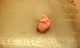


126 a b Figure 11. Root surfaces after root-end resections. Complete isthmus (a) and partial isthmus (b) between mesiobuccal and mesiolingual root canals (stereomicroscope x30). a b Figure 12. Root surfaces after root-end resections. Complete isthmus (a) and partial isthmus (b) between mesiobuccal and mesiolingual root canals (SEM x40). 65


")
,")
 and c) SEM")


 (x63)")


127 a b c Figure 13. Observation of the same root surface under three magnification methods: a) operative microscope (x16), b) stereomicroscope (x30) and c) SEM (x30). A complete isthmus was visible only with SEM, while with operative and stereomicroscope was misinterpreted as partial isthmus. a b Figure 14. Observation of the same root surface under stereomicroscope (a) (x63) and SEM (b) (x30). Complete isthmus misinterpreted as partial isthmus under stereomicroscope ). 66

128 a b Figure 15. Observation of the same root surface under stereomicroscope (a) (x63) and SEM (b) (x30). No isthmus present under stereomicroscope, whereas a partial isthmus was observed under SEM. The age related variable and its correlation with the prevalence of different types of isthmuses were recorded but not further analyzed in this study, as the sample was relatively small to fulfill the requirements of morphological / anatomical studies to be interpretable and reliable. As such, no extrapolation methods were used. 67
129 4.3 Number Type of microcracks Stereomicroscope and SEM were used to examine the presence and type of microcracks. Of the 60 mesial roots observed before root end preparations under stereomicroscope, 8 displayed incomplete type A microcracks, 1 had an incomplete type B and 2 had complete microcracks, regardless the retrotip used. After the root end preparations the roots displayed 10, 2 and 5 microcracks, respectively. These findings correspond to the formation of 2 new incomplete type A, 1 new incomplete type B and 3 new complete microcracks (Figure 16). Of the 60 mesial roots observed before root end preparations under SEM, 20 displayed incomplete type A microcracks, 3 had an incomplete type B and 2 had complete microcracks, regardless of the retro tip used. After the root end preparations the roots displayed 25, 5 and 7 microcracks, respectively. These findings correspond to the formation of 5 new incomplete type A, 2 new incomplete type B and 5 new complete microcracks (Figure 16). 68

")
.")
")


130 a b c Figure 16. Comparison of same surfaces under stereomicroscope (a) (x30) and SEM (b, c) (x30). Two incomplete type A microcracks (squares) were identified with both magnification methods but one incomplete type A (arrow) was observed only under SEM (c). 69
131 A total number of 11 microcracks before the root end preparations were identified under stereomicroscope and 25 microcracks under SEM. After the root end preparations these numbers were 17 and 37 respectively, meaning to that 14 microcracks before and 20 microcracks after the root end preparations were not detected by the stereomicroscope. For the sake of agreement we assume that the sensitivity is 100% of SEM accuracy regarding the identification of microcracks. As such the stereomicroscope revealed 44% and 46% sensitivity before and after the root end preparations. All previous data are listed in tables 3 & 4. A statistical significant difference was found between stereomicroscope and SEM when the total number of microcracks was compared before and after the root end preparations (P<0.05, chi square test). The intra observer reliability was estimated after 2 week time intervals and interpretation for Cohen s κ value amounted 0.47, corresponding to a moderate agreement. The value was calculated by repeated observations under stereomicroscope. 70
132 Table 3. Distribution of microcracks before and after root-end preparations with two magnification methods. Incomplete type A (n=60) Incomplete type B (n=60) Complete (n=60) before after before after before after Stereomiroscope SEM Significant difference between stereomicroscope and SEM with regard to the total number of microcracks detected (P<0.05, chi square test). Table 4. Total number of microcracks before and after root-end preparations and sensitivity of magnification methods Total number of microcracks (n=60) Sensitivity (%) before after before after Stereomicroscope SEM The distribution of the number and type of microcracks among the three groups of different retrotips is summarized in table 5. The main evaluation, performed under SEM, showed the following results: 71
133 Group A Of the 20 roots observed before root end preparations, 6 roots had 9 incomplete type A microcracks, 1 root had 2 incomplete type B and 1 root had a complete microcrack (total number of microcracks: 12). After root end preparations, 7 roots had 11 incomplete type A (2 new incomplete type A) and 2 roots had 3 complete microcracks (2 new complete). There was no new formation of incomplete type B microcrack (total number of microcracks: 16). There was no change in length or width in any pre existing microcrack after root end preparations. There were 3 microcracks situated around the isthmus area. No overall microcrack formation was recorded in 12 roots before and 10 after the root end preparations. Group B Of the 20 roots observed before root end preparations, 6 roots had 6 incomplete type A microcracks and no roots had incomplete type B or a complete microcrack (total number of microcracks: 6). After root end preparations, 6 roots had 7 incomplete type A (1 new type A), 1 root had an incomplete type B (1 new type B) and 1 root had 2 complete microcracks (total number of microcracks: 10). One complete microcrack increased in width after root end preparations. There was no microcrack around the isthmus area. No overall 72
134 microcrack formation was recorded in 14 roots before and 12 after the root end preparations. Group C Of the 20 roots observed before root end preparations, 5 roots had 5 incomplete type A microcracks, 1 root had a incomplete type B and 1 root had a complete microcrack (total number of microcracks: 7). After root end preparations, 5 roots had 7 incomplete type A (2 new type A), 1 root had 2 incomplete type B (1 new type B) and 2 roots had 2 complete microcracks (total number of microcracks: 11). An incomplete type A increased in length after root end preparations. There were 3 microcracks observed around the isthmus area. No overall microcrack formation was recorded in 13 roots before and 12 after the root end preparations (Figures 17 24) Statistical analysis showed no significant difference on the incidence of microcracking when three retrotips were compared (P>0.05, Kruskal Wallis U test). There was no significant differences in the type of microcracks produced by the retrotips (P>0.05, Kruskal Wallis U test). Finally, a significant larger number of microcracks was observed around the root canals than these observed around the isthmuses (P<0.05, chi square test). 73
135 The intra observer reliability was estimated after 2 week time intervals and interpretation for Cohen s κ value amounted 0.88, corresponding to an almost perfect agreement. The value was calculated by repeated observations on the SEM images taken on the first round. Table 5. Microcrack distribution before and after root-end preparations under SEM evaluation. group A (n=20) group B (n=20) group C (n=20) before after before after before after Incomplete type A Incomplete type B Complete Total Group A: diamond coated retrotips (P14 D), group B: diamond coated retrotips (KiS 1), group C: zirconium nitride coated (Surg 1). No Significant difference between three groups with regard to the number and type of microcracks (P>0.05, Kruskal Wallis U test). 74

.")

136 a b c Figure 17. Comparison of the same root surface before and after root-end preparation with P14-D (group A). a) before: no microcrack, b) after: new incomplete type A (x30 magnification), c) (x80 magnification) 75

 before: incomplete type A microcrack, b) after: pre-existing")
137 a b Figure 18. Comparison of the same root surface before and after root-end preparation with KiS-1 (group B). a) before: incomplete type A microcrack, b) after: pre-existing incomplete type A and formation of a new incomplete type A (x30 magnification). 76

 before: no microcrack, b) after: new")
138 a b Figure 19. Comparison of the same root surface before and after root-end preparation with Surg-1 (group C). a) before: no microcrack, b) after: new incomplete type A (x30 magnification). 77

 before: incomplete type A microcrack, b) after: the")
139 a b Figure 20. Comparison of the same root surface before and after root-end preparation with Surg-1 (group C). a) before: incomplete type A microcrack, b) after: the same pre-existing incomplete type A (x80 magnification). 78

 before: incomplete type B microcracks, b) after: the")
140 a b Figure 21. Comparison of the same root surface before and after root-end preparation with P14-D (group A). a) before: incomplete type B microcracks, b) after: the same pre-existing incomplete type B (x30 magnification). 79

.")
141 a b Figure 22. Comparison of the same root surface before and after root-end preparation with P14-D (group A). a) before: no microcrack, b) after: new complete (x30 magnification). 80

 before: complete microcrack, b) after: the same")
142 a b Figure 23. Comparison of the same root surface before and after root-end preparation with Surg-1 (group C). a) before: complete microcrack, b) after: the same pre-existing complete (x35 magnification). 81

.")
143 a b Figure 24. Comparison of the same root surface before and after root-end preparation with Kis-1 (group B). No microcrack observed before (a) and after (b) (x30 magnification). 82
144 4.4 Marginal chipping The results of the evaluation under SEM of the root end preparations, with regard to marginal chipping are summarized in table 6. Group A Of the 20 roots observed after root end preparation, 9 root end preparations had no chipping, 8 had microchipping and 3 had chipping. None was detected with imprint formation (total score: 25). Group B Of the 20 roots observed, 15 had no chipping, 2 had imprint, 3 had microchipping and 1 had chipping (total score: 11). Group C Of the 20 roots observed, 16 had no chipping, 1 had imprint, 4 had microchipping and 1 had chipping (total score: 12). These findings derived after capturing three visual fields in every specimen (x30 magnification), one for each root canal and one for the isthmus area, forming a total of 60 images for each group (Figure 25). 83
145 In a similar way as for microcracks, the intra observer reliability was estimated after 2 week time intervals and interpretation for Cohen s κ value amounted 0.79, corresponding to a substantial agreement. The value was calculated by repeated observations on the SEM images taken on the first round. Following Kruskal Wallis U tests, it appeared that all ultrasonic retrotips in the three groups produced overall marginal chipping (P>0.05). There was no significant difference in the number of root end preparations without chipping among the groups (P>0.05). 84
146 Table 6. Marginal chipping formation after root-end preparations. group A (n=20) group B (n=20) group C (n=20) No chipping (score O) Imprint (score 1) Microchipping (score 2) Chipping (score 3) Total (total score) 25* 11** 12*** * Total score in group Α: (8x2)+(3x3)=25, ** group Β: (2x1)+(3x2)+(1x3)= 11, *** group C: (1x1)+(4x2)+(1x3)= 12. No significant difference between the three groups with regard to the marginal chipping formation (P=0.0975, Kruskal Wallis U test). 85


 no")

 . (x30,")
147 a b c d e f Figure 25. Marginal chipping after root-end preparations. a) no marginal chipping, b) imprint, c, d) microchipping, e, f) chipping. (x30, SEM). 86
148 4.5 Time recording The mean and the standard deviation of the root end preparation time for groups A, B and C was 2.48 ± 0.41, 1.53 ± 0.38, 1.57 ± 0.43, respectively (Table 7). More time was required for group A after statistical comparison of the values of the three root end preparations (P<0.01, one way ANOVA test). No further analysis and correlation was done between the prevalence or the type of isthmuses and the preparation time in the current study. Table 7. Mean root-end preparation time (min) among the three groups. group A group B group C (Mean ± SD) 2.48 ± 0.41 a 1.53 ± ± 0.43 a Significant difference between retrotips overall root end preparation time (P<0.01, one way ANOVA test). 87
149 5. Discussion For over ten years, the use of ultrasonic retrotips has been enabling the most effective clinical protocols in the field of surgical endodontics, being established as an inextricable instrument in the surgeons armamentarium. Recent improvements in their characteristics facilitate adequate root end preparations of narrow, difficult to access and prepare, isthmuses of the root canal system. The objective of the present ex vivo study was to investigate the incidence of root canal isthmuses in the mesial root of mandibular first molars after root end resections, using different magnification methods. A second fold was to compare the effect of three, recently available, two diamond and one zirconium nitride coated ultrasonic retrotips on the root surfaces of the mesial root of mandibular first molars. 5.1 Discussion of method Root canal treatment Extensive manipulation during orthograde endodontic treatment and subsequent modification in the root canal system morphology may cause an 88
150 increased number of dentin microcracks (Morgan & Marshall 1999, Gondim et al. 2002). Beling et al. (1997) pointed out insignificant difference between root canal treated and uninstrumented teeth, with regard to the number and type of microcracks. In the present study, root canal treatment was performed, mimicking clinical conditions. To avoid any misinterpretation the teeth were inspected under stereomicroscope and this potential was, also, considered when evaluating the root surfaces after the resection of the apices under scanning electron microscope (SEM) Artificial periodontal model As stated in previous studies (Taschieri et al. 2004, De Bruyne & De Moor 2005), the absence of periodontal ligament may influence the formation of microcracks as no normal surrounding tissues are present to absorb the total ultrasonic energy. The possible absence of energy absorption of ultrasonic vibrations in the periodontal ligament and the alveolar bone might increase the mechanical transference of energy to the root surface and produce more artifactual microcracks and marginal chipping during ultrasonic root end preparations (Taschieri et al. 2004, De Bruyne & De Moor 2005). Efforts to simulate the periradicular tissues by using an acrylic resin block (Khabbaz et al. 2004) and to 89
151 stabilise the teeth during the apical root end procedures by using a fixing apparatus (Gondim et al. 2002), were used in previous experiments. In the present study an artificial periodontal model has been developed out of a polyvinylsiloxane impression material and a fixing apparatus were used, in order to achieve more precise simulation of the clinical conditions (Peters et al. 2001, Dobo Nagy et al. 2003, Soares et al. 2005). Any comparison between teeth with or without the application of artificial periodontal model and fixing apparatus was not part of the current experiment and was not further evaluated. The influence of periradicular tissues in the absorbance of the ultrasonic vibrations is further supported by the findings of studies in human cadavers. De Bruyne & De Moor (2005) concluded that cadaver teeth had fewer microcracks and chipping than the extracted teeth. Two former studies comparing human cadavers and extracted teeth in regard to microcracking and chipping potential, found no microcrack and chipping formation and no statistical significant difference between cadavers and extracted teeth, respectively (Calzonetti et al. 1998, Gray et al. 2000). The difference in the results of these studies did not confirm that the presence of periradicular tissues account for the decreased formation of microcracks and marginal chipping. Other related factors should be evaluated comprehensively. Nevertheless, the 90
152 application of artificial periodontal model or presence of periradicular tissues when possible, are considered to be beneficial as they mimic the clinical conditions Root-end resection The apical 3mm of each mesial root was resected perpendicular to the long axis of the root using a high speed tungsten carbide cylindrical bur. The selection of bur was based on an ex vivo experimental protocol from Weston et al. (1999). After comparing several rotary cutting burs they concluded that no significant disruption, distortion or smearing of the gutta percha and the sealer was occurred when burs were used in the forward direction. The tungsten carbide cylindrical bur produced a surface with parallel grooves and ridges on all the root surfaces, which could be inspected under SEM, similar to the images produced in the current study. The extent and the bevel angle of the apical resection in this study were based on the most current principles in surgical endodontics (Kim et al. 2001, 2006, Bradford & Witherspoon 2006). 91
153 5.1.4 Parameters of ultrasonic generators Both retrotips and piezo electric ultrasonic generators are available from various manufactures, operating under variable settings. Previous studies used low, medium or high power modes, corresponding to different frequencies and amplitudes. The standardization of these parameters and their effect on the overall performance of different retrotips generates a number of controversial viewpoints. In a recent study, Thomson et al. (2007) investigated the vibration characteristics of a retrotip (KiS 2; Spartan Obtura, Fenton, USA) through the evaluation of variable power modes in different ultrasonic generators and handpieces, using a scanning electron vibrometer. The authors concluded that there was no linear increase in vibration displacement amplitude of the retrotip with increase from low to medium power in the generators tested. Different types of retrotips and generators need to be tested in the future. Additionally, they found variability in power output between generators and handpieces of the same manufacturer when compared with each other. In the present study, the same piezo electric ultrasonic generator and handpiece was used for all root end preparations, according to the aforementioned findings. According to the manufactures brochure the new generation units are controlled by unique electronic module, operating at frequency khz, 92
154 power range W, vibration amplitudes 4μm 200μm (ISO 22374:2005 standard requirements), parameters that were not fully standardized in the present generator. Nevertheless, the power was set in mode E, scale 5 7, that equals to medium power, following the interrelation between older and newer protocols, mentioned in the manufactures official brochure. The power mode S corresponds to high power and is not recommended by the updated protocols from the manufacturer, although it has been used effectively in several studies in the past in combination with different type of retrotips (De Bruyne & De Moor 2005). With regard to microcrack formation several studies have been performed in the past testing different power settings (Frank et al. 1995, Layton 1996, Ishikawa et al. 2003, Taschieri et al. 2004). Based on their results, both high and low power settings can contribute to the formation of microcracks, producing different amplitudes. In the present study, medium power settings were used for all the root end preparation, following the manufactures recommendations and the findings of previous studies (Taschieri et al. 2004, De Bruyne & De Moor 2005). 93
155 5.1.5 Methodology of root-end preparation The root end preparation methodology for all tested retrotips proposed in this study is a modification of the previously described methods (Carr 1997, Kim 2001). Starting the preparation from one root canal allows the operator to locate better the final retrograde cavity. Kim (2001) suggests the orientation of the root end preparation should be from buccal to lingual. This variable depends, mainly, on the available operation field during clinical conditions, assuming that the mesiobuccal root canal is easier to be located and can act as a guide to the remaining root end preparation. According to the methodology followed in the current study, the first stage of isthmus preparation using a lower power (scale 2 3) and a micro explorer for tracking enabled a well orientated and controlled preliminary preparation. The possibility of perforations in the roots, breakage of the retrotips and formation of less crazing (scratches and grooves) on the root surfaces by avoiding the misplacement of the retro tips, was eliminated SEM parameters The preparation of the samples for SEM evaluation is of major importance, in order to choose a non destructive technique that allows a comparison of the 94
156 samples before and after the root end preparations, and to exclude the formation of artifactual cracking, which may result in misinterpretation and overestimation of the final data. In the present study a significant account was taken in order to decide the appropriate samples preparation and overcome procedural artifacts. A preliminary pilot study was performed to choose an appropriate nondestructive preparation method for LV SEM evaluation and to standardize the critical parameters that can influence the results. It was based on the experimental methods for dry uncoated and coated specimens used by Shipper et al. (2004) and indirect replica technique used in a previous study (Beltes 2006). In the present study, all specimens were examined under the same low vacuum conditions accelerating potential of 10kV and total pressure between Torr, values that enable elimination of the critical point drying. The direct method of the specimens which involved desiccation and coating was not preferred, as this method causes possible alteration on the root surfaces, which does not allow the comparison of the same specimen before and after the rootend preparation under the same magnification device. Considering the inevitable existing artifactual potential in all experimental in vitro and ex vivo studies, the present study applied the direct dry non coated 95
157 preparation method proposed by Shipper et al. (2004) and supported by the current pilot study. To the best of our knowledge, this is the first time in the literature that such a method is used for direct comparison of root surfaces before and after root end preparations. 5.2 Discussion of results Prevalence of isthmuses The identification of isthmuses is of critical importance, as these anatomical structures can act as reservoir for accumulation of microorganisms, necrotic tissue or can act as an exit portal of an unsealed part of the root canal system. As such, isthmuses can be a reason for non healing process of the periradicular pathology after conventional and surgical endodontics (Hsu & Kim 1997, Texeira et al. 2003). In general, this anatomical complexity remains inadequately instrumented, irrigated and obturated during root canal treatment and conditions the prognosis of root canal treatment (Nair et al. 2005). Similar principles apply to the prognosis of periradicular surgery, respectively. The classification for isthmuses used in previous studies was based on the findings of Hsu & Kim (1997). The authors identified five types, in which they 96
158 included the presence of a third root canal (mesial medial root canal). In the present study, a third root canal was not identified in any of the specimens during root canal treatment or after 3mm resection of the apex, thereby this classification was subjective. This finding is justified because of the very low incidence of a third root canal in the mesial root or the unification in the apical third when present (Fabra Campos 1989). The classification applied was similar to Mannocci et al. (2005), dividing the findings to three categories: a) absence of isthmus, when there was no notable intercanal communication, b) partial isthmus, when there was definite or, even, a barely traceable communication and c) complete isthmus, when there was a continuous opening between two root canals. Based on the above classification, the incidence of isthmus formation under SEM evaluation in this study, without reference to the type, was almost 100% (59/60). Incongruity of the results with previous studies (Hsu & Kim 1997, Teixeira et al. 2003, von Arx et al. 2005, Mannocci et al. 2005), which shown a prevalence of 50 83%, can be related to contributing factors, such as the number of samples, magnification methods and chosen classification. In the present study traceable communications and thin discontinuations of dentin between two mesial root canals were classified as partial isthmuses, observations that can, possibly, increase the total percentage of isthmuses. 97
.")
 (Figure 26) Figure 26.")
159 With regard to the type of isthmuses, the occurrence of partial isthmuses was found to be more frequent than complete isthmuses, similar to the findings of previous studies (Weller et al. 1995, Hsu & Kim 1997, Teixeira et al. 2003, Mannocci et al. 2005). All isthmuses showed unsealed areas between the root canal sealer, gutta percha and dentine walls, regardless the type of the isthmus, observations that are in agreement with other authors and emphasize the necessity of preparation and hermetic sealing of these anatomical structures (Peters et al. 2003, Nair et al. 2005) (Figure 26) Figure 26. Unsealed isthmus areas observed under stereomicroscope (x30) and SEM (x40) In the attempt to evaluate the accuracy of different magnification methods, in isthmus identification, a SEM was used as a golden standard method to examine surfaces and biomaterials and compared to an operative microscope 98
160 and a stereomicroscope. In terms of sensitivity, the operative microscope revealed 73% and the stereomicroscope 90%, while the SEM had a standard variable of 100%. The overall low ability of the operative microscope to identify isthmuses in this study could have been influenced by the indirect examination through a micro mirror, the limited field of observation as teeth were placed on an apparatus and, mainly, the absence of staining application (Stropko 1999, Kim 2002). Stains were not used in this study to avoid alterations of the surfaces which may affect the next stages of the experiment. The use of stereomicroscope determined better visual inspection but when compared with SEM was found less accurate. The null hypothesis (H0) in this study stated that different magnification methods can detect with the same accuracy the prevalence of isthmuses, is rejected. It can be concluded that examinations performed with these methods may lead to misinterpretation of the number and type of isthmuses and give different results among studies. Therefore, high resolution and magnification devises, like SEM (LV SEM or E SEM if uncoated specimens are used) should be preferred for a better standardization of the observational parameters. In recent years, high resolution micro computed tomography (mct) was used to evaluate morphology of root canal system or to compare root preparation and obturation techniques, as it allows analysis without necessitating sectioning of 99
161 the specimens (Peters et al. 2003, Loizides et al. 2007). This costly and, also, time consuming method was used to detect the presence of isthmuses for the first time by Mannocci et al. (2005) and can be an auspicious non destructive innovation in the future. The dilemma among surgeons arises when the presence of an isthmus is not identified under operative microscope in clinical conditions, but might exist when examined under SEM or stereomicroscope. Supported by the findings of several ex vivo studies, including the present one, when two root canals are located the presence of an isthmus should be expected and included in the rootend preparation and retrograde obturation. There are no randomized controlled clinical studies investigating the healing outcome of prepared and sealed versus unprepared isthmuses in the field of surgical endodontics and their causal relationship should be clarified. Furthermore, the extent to which this reflects the formation of microcracks is unclear and any possible correlation between the presence, type of isthmuses and number of microcracks will be further analyzed in the following chapter. 100
162 5.2.2 Microcrack formation During the last decade ultrasonic root end preparation techniques have gained increased popularity in the field of periradicular surgery. The advantages when compared with conventional round burs have been well demonstrated including, easier access to the cavities, preparation of deeper retrograde cavities with parallel walls that follow the original anatomy of the root canal and decreased possibility of root perforation. They produce less smear layer, less exposure of dentinal tubules and remove less dental hard tissue, contributing to better retention of the retrograde filling materials (Abedi et al. 1995, Engel & Steiman 1995, Carr 1997, Lin et al. 1998). The use of ultrasonic retrotips provides superior control and preparation of thin anatomical structures, such as isthmuses, without extensive manipulation avoiding iatrogenic complications. On the contrary, Saunders et al. (1994) reported that the use of ultrasonic retrotips can be a predisposing factor for the formation of microcracks in the root surface. The significance of microcracking has been suggested to increase the susceptibility to root fracture, apical microleakage of retrograde filling material and increase the possibility of bacterial contamination (Saunders et al. 1995, Gray et al. 2000). It has been stated that existing microcracks in the root surface can increase in size after retrograde obturation and loading conditions, causing microleakage (Saunders et al. 1995). Whether these factors result in 101
163 fracture formation or bacterial accumulation during functioning, with an overall non healing outcome, should be further investigated (Peters & Peters 2002, Tobon Arroyave et al. 2007). In the present study, a SEM and a stereomicroscope were compared, in order to evaluate the accuracy of different magnification methods, with regard to microcrack identification, Assuming that the SEM has a standard variable of 100% sensitivity, the stereomicroscope revealed 44% sensitivity before and 46% after the root end preparations. There was a significant difference in the number of incomplete type A and B microcracks detected under the two devices. These findings suggest that the stereomicroscope was not able to detect more than half of the existing microcracks, especially in terms of microcracks with smaller dimensions like incomplete type A and B. The null hypothesis (H0) that different magnification methods can detect with the same accuracy the prevalence and type of microcracks is rejected. Results of previous studies using additional staining to enhance visualization under stereomicroscope can t, therefore, be compared with this study. In contrast, Engel & Steiman (1995) although using methylene blue in conjunction with stereomicroscope found a significant smaller number of microcracks. These findings can be related to the lower magnification (x20) used in that study. 102
164 Generally, this variation in accuracy may lead in misinterpretation between the findings and result in false positive or negative conclusions. The occurrence of microcracks before the root end preparation was not taken under consideration in the past, which resulted in an inaccurate estimation of the incidence of microcrack formation after root end preparation. Any microcrack formation can be produced during root end resection, extractions, orthograde root canal treatment or can be related to the aging processes (Layton et al. 1996). In the present study, an initial microcrack incidence of 41.7% (25/60) was found after root end resection, a finding that corresponds with those of other investigators (Abedi et al. 1995, De Bruyne & De Moor 2005, Beltes 2006) who have reported an incidence of 28%, 35,5%, 26,7% respectively. The relatively higher percentage of microcracraks in this study can be associated with the different evaluation method and the group of teeth, as all the previous studies were performed in single rooted teeth. However, the present study is in agreement with the previous studies (Gondim et al. 2002, Taschieri et al. 2004) regarding the type of microcracks present before and after the root end preparations, forasmuch as incomplete type A microcrack being the most common. The larger number of incomplete type A microcracks might be related to the instrumentation and obturation procedures during orthograde 103
165 endodontic treatment and the vibration effect of the retrotips during root end preparations. In the present study, the amount of microcracks was higher than two of the previous studies that used molars (Engel & Steiman 1995, Peters et al. 2001) but variables like the number of teeth, the observation method and the absence of comparison before and after the root end preparation can alter the results. The incidence of microcracks in this study is supported by the findings in other studies, which used either sensitive evaluation methods (Min et al. 1997), or direct and indirect preparation for SEM evaluation (Abedi et al. 1995, Calzonetti et al. 1998, Taschieri et al. 2004, De Bruyne & De Moor 2005). In the present study, 5 new complete microcracks were observed regardless of the type of retrotip used, corresponding to 8% of total microcrack incident. These results are similar to those of previous studies (Abedi et al. 1995, De Bruyne & De Moor 2005, Beltes 2006). Alterations in the length and width of pre existing microcracks after the rootend preparations were noticed in 2 out of 25 microcracks. These findings suggest that the ultrasonic retrotips used in this study did not have a direct impact in the microcracks existing before root end preparations, whereas in previous studies the results were contradictory (Rainwater et al. 2000, Beltes 2006). Contributing factors might be the presence of artificial periodontal model 104
166 and apparatus, resulting in a better absorption of the overall vibration and stability, respectively. Another objective of this study was to assess if different types of coated retrotips produced different number and type of microcracks or altered the size of pre existing microcracks. Two studies compared diamond and zirconium nitride coated retrotips (Navarre & Steiman 2002, Ishikawa et al. 2003). Navarre & Steiman (2002) found no microcrack formation in both types after evaluating 50 single rooted teeth under operative microscope. Under similar observation methods, Ishikawa et al. (2003) found a total of 3 microcracks in 30 single rooted teeth, without specifying their type. To the best of our knowledge, the present study is the first in the literature comparing thinner types of diamond and zirconium nitride coated retrotips and, also, the first comparing these types of retrotips in the mesial root of the first mandibular molar under SEM. In the current study, all retrotips tested produced new microcracks. A total of 12 new microcracks were observed without any significant difference between the type of microcracks. This is 20% (12/60) of the overall microcrack formation, present in 26 out 60 roots. The remaining 34 roots were free of microcracks after the completion of the experiment. These findings are in agreement with previous studies, using non destructive techniques to compare root surfaces before and after the root end preparations (Abedi et al. 1995, Beling et al. 1997, Rainwater et 105
167 al. 2000, De Bruyne & De Moor 2005, Beltes 2006). Therefore, the type of retrotip tested, seemed not to play a role in the formation of microcracks. The formation of microcracks with any type of retrotip is an incident to be anticipated and, although controversial, may be an important factor that might influence the healing process of the periradicular tissues. Further clarification is needed, both in ex vivo and in vivo studies, to prove that microcracks can cause failure of periradicular surgery, even if the overall number is small. Until proven otherwise, we should attempt to minimize the number or seal the existing microcracks. The possible benefits of mechanical removal of such defects by finishing and burnishing resected surfaces after the end of the retrograde obturation has been investigated, but microleakage was not avoided (Gondim et al. 2005). In terms of biomaterials, Rud et al. (1991, 1996, 2001) in several studies reported that sealing of a concaved resected root end with a dentine bonding agent and a flowable resin composite (Gluma Retroplast ) will result in the prevention of microleakage, by covering all anatomical structures including isthmuses, accessory canals, ramifications, anastomoses and possible microcracks. Longterm prospective clinical studies using this retrograde filling material reported an overall successful healing of more than 90% with follow up period ranging 106
168 from 1 to 12 years (Rud et al. 2001, Jensen et al. 2002, Yazdi et al. 2007). Moreover, Sarkar et al. (2005) demonstrated that mineral trioxide aggregate (MTA), apart from the, well documented, positive effect on cementum deposition and osseous healing (Torabinejad et al. 1995) has the ability to precipitate hydroxyapatite (HA) micro crystals when phosphate buffered saline is present, which may be relevant in minimizing microleakage thereafter. Randomized clinical studies in the literature that used MTA as a retrograde filling material reported an overall 90% successful healing (Chong et al. 2003, Saunders 2008). Any relevance between microcracking and clinical outcome should be further investigated. The effect of ultrasonic retrotips in the remaining dentine around the isthmus area was also evaluated in this study, with regard to the formation of microcracks. The results suggested no association between thin dentin and microcrack formation, regardless of the type of retrotip used. These findings concur with those reported in previous studies (Gondim et al. 2002, Khabbaz et al. 2004), which used single rooted teeth of different dimensions. Other authors reported opposite results and correlated thin dentin surfaces, less than 1mm, with microcracking potential (Abedi et al. 1995, Frank et al. 1996). Although the former results are not supported, it is prudent to use ultrasonic retrotips conservatively when a narrow, biconcaved root is present. The biconcave 107
169 middle portion of a mandibular mesial or a mesio buccal root of a maxillary molar can be potentially over prepared and weakened after root end preparation, predisposing to iatrogenic complications and failure of periradicular surgery (Peters et al. 2002). The use of retrotips with small diameter in this study prevented any unnecessary removal of the surrounding dentin and any perforation was avoided, incident that was recorded in a previous study (Peters et al. 2002). Following the root end methodology proposed in this study, there was no perforation in any of the roots, even in very narrow regions around the isthmuses (Figure 27). Crazing was observed under SEM in 5 roots of group A with partial isthmuses. This finding can be explained by the fact that P14 D retrotip is larger by 0.1mm in diameter and more rounded at the end of the cutting edge (tip), characteristics that predispose a possible sliding on the root surface during the root end preparation (Figure 28). In order to eliminate these defects some authors suggested a final burnishing of the root surface and the root end filling material (Peters et al. 2001). The null hypothesis (H0) of equivalence after root end preparations with three different ultrasonic retrotips regarding the formation of microcracks is accepted. 108


170 Figure 27. Root-end preparation of isthmuses in narrow biconcaved roots (x30). Perforation was avoided due to application of thin ultrasonic retrotips. Figure 28. Sliding of P14-D retrotip (group A) during root-end preparation (x30) 109
ΔΙΑΝΟΙΞΗ ΓΙΑ ΕΝΔΟΔΟΝΤΙΚΗ ΘΕΡΑΠΕΙΑ. ΣΤΟΧΟΙ, ΜΕΣΑ ΚΑΙ ΣΥΜΒΑΜΑΤΑ.
 ΕΡΓΑΣΤΗΡΙΟ ΕΝΔΟΔΟΝΤΙΑΣ ΟΔΟΝΤΙΑΤΡΙΚΗ ΣΧΟΛΗ ΠΑΝΕΠΙΣΤΗΜΙΟΥ ΑΘΗΝΩΝ 3 Ο ΕΡΓΑΣΤΗΡΙΑΚΟ ΣΕΜΙΝΑΡΙΟ ΕΝΔΟΔΟΝΤΙΑΣ 5 ΟΥ ΕΞΑΜΗΝΟΥ ΔΙΑΝΟΙΞΗ ΓΙΑ ΕΝΔΟΔΟΝΤΙΚΗ ΘΕΡΑΠΕΙΑ. ΣΤΟΧΟΙ, ΜΕΣΑ ΚΑΙ ΣΥΜΒΑΜΑΤΑ. ΣΚΟΠΟΣ ΤΟΥ ΣΕΜΙΝΑΡΙΟΥ
ΚΛΑΣΣΙΚΗ ΚΑΙ ΣΥΓΧΡΟΝΗ ΕΝΔΟΔΟΝΤΙΑ: ΑΠΟ ΤΗ ΘΕΩΡΙΑ ΣΤΗΝ ΚΛΙΝΙΚΗ ΠΡΑΞΗ
 ΚΛΑΣΣΙΚΗ ΚΑΙ ΣΥΓΧΡΟΝΗ ΕΝΔΟΔΟΝΤΙΑ: ΑΠΟ ΤΗ ΘΕΩΡΙΑ ΣΤΗΝ ΚΛΙΝΙΚΗ ΠΡΑΞΗ Υπεύθυνος δραστηριότητας: Μ. Γ. ΧΑΜΠΑΖ Σκοπός: Σκοπός αυτού του μετεκπαιδευτικού προγράμματος είναι η συνεχής επιμόρφωση των οδοντιάτρων
ΕΡΓΑΣΤΗΡΙΟ ΕΝΔΟΔΟΝΤΙΑΣ. ΟΔΟΝΤΙΑΤΡΙΚΗ ΣΧΟΛΗ ΠΑΝΕΠΙΣΤΗΜΙΟΥ ΑΘΗΝΩΝ 5 Ο ΕΡΓΑΣΤΗΡΙΑΚΟ ΣΕΜΙΝΑΡΙΟ ΕΝΔΟΔΟΝΤΙΑΣ 5 ΟΥ ΕΞΑΜΗΝΟΥ ΥΠΟΛΟΓΙΣΜΟΣ ΤΟΥ ΜΗΚΟΥΣ ΕΡΓΑΣΙΑΣ
 ΕΡΓΑΣΤΗΡΙΟ ΕΝΔΟΔΟΝΤΙΑΣ. ΟΔΟΝΤΙΑΤΡΙΚΗ ΣΧΟΛΗ ΠΑΝΕΠΙΣΤΗΜΙΟΥ ΑΘΗΝΩΝ 5 Ο ΕΡΓΑΣΤΗΡΙΑΚΟ ΣΕΜΙΝΑΡΙΟ ΕΝΔΟΔΟΝΤΙΑΣ 5 ΟΥ ΕΞΑΜΗΝΟΥ ΥΠΟΛΟΓΙΣΜΟΣ ΤΟΥ ΜΗΚΟΥΣ ΕΡΓΑΣΙΑΣ ΣΚΟΠΟΣ ΤΟΥ ΣΕΜΙΝΑΡΙΟΥ Είναι να αποκτήσουν οι φοιτητές
Αντιμετώπιση δοντιών με εκτεταμένη απώλεια ιστών
 Στρατής Παπάζογλου Προσθετολόγος, MS, PhD Επικ. Καθηγητής Οδοντικής Χειρουργικής Αντιμετώπιση δοντιών με εκτεταμένη απώλεια ιστών Εκτεταμένη απώλεια ιστών Κοιλότητες μεγαλύτερες από τυπικές, συχνά αποκαθυστούν
ΕΛΛΗΝΙΚΗ ΕΝΔΟΔΟΝΤΙΚΗ ΕΤΑΙΡΕΙΑ. HELLENIC SOCIETY of ENDODONTICS. Ενδοδοντική θεραπεία. Τι θα πρέπει να γνωρίζουμε
 ΕΛΛΗΝΙΚΗ ΕΝΔΟΔΟΝΤΙΚΗ ΕΤΑΙΡΕΙΑ HELLENIC SOCIETY of ENDODONTICS Ενδοδοντική θεραπεία Τι θα πρέπει να γνωρίζουμε Ενδοδοντική θεραπεία Τι θα πρέπει να γνωρίζουμε Το δόντι αποτελείται από σκληρούς ενασβεστιωμένους
ΣΥΜΒΑΜΑΤΑ ΚΑΤΑ ΤΗ ΧΗΜΙΚΟΜΗΧΑΝΙΚΗ ΕΠΕΞΕΡΓΑΣΙΑ ΤΩΝ ΡΙΖΙΚΩΝ ΣΩΛΗΝΩΝ.
 ΟΔΟΝΤΙΑΤΡΙΚΗ ΣΧΟΛΗ ΠΑΝΕΠΙΣΤΗΜΙΟΥ ΑΘΗΝΩΝ. ΕΡΓΑΣΤΗΡΙΟ ΕΝΔΟΔΟΝΤΙΑΣ. 7 Ο ΕΡΓΑΣΤΗΡΙΑΚΟ ΣΕΜΙΝΑΡΙΟ ΕΝΔΟΔΟΝΤΙΑΣ 5 ΟΥ ΕΞΑΜΗΝΟΥ ΣΥΜΒΑΜΑΤΑ ΚΑΤΑ ΤΗ ΧΗΜΙΚΟΜΗΧΑΝΙΚΗ ΕΠΕΞΕΡΓΑΣΙΑ ΤΩΝ ΡΙΖΙΚΩΝ ΣΩΛΗΝΩΝ. Σκοπός του Σεμιναρίου:
ΣΥΜΒΑΜΑΤΑ ΚΑΤΑ ΤΗΝ ΕΜΦΡΑΞΗ ΤΩΝ ΡΙΖΙΚΩΝ ΣΩΛΗΝΩΝ
 ΟΔΟΝΤΙΑΤΡΙΚΗ ΣΧΟΛΗ ΠΑΝΕΠΙΣΤΗΜΙΟΥ ΑΘΗΝΩΝ. ΕΡΓΑΣΤΗΡΙΟ ΕΝΔΟΔΟΝΤΙΑΣ. 10 ο ΕΡΓΑΣΤΗΡΙΑΚΟ ΣΕΜΙΝΑΡΙΟ ΕΝΔΟΔΟΝΤΙΑΣ 5 ΟΥ ΕΞΑΜΗΝΟΥ ΣΥΜΒΑΜΑΤΑ ΚΑΤΑ ΤΗΝ ΕΜΦΡΑΞΗ ΤΩΝ ΡΙΖΙΚΩΝ ΣΩΛΗΝΩΝ Σκοπός του Σεμιναρίου: Είναι να γνωρίσουν
ΟΔΟΝΤΙΑΤΡΙΚΗ ΣΧΟΛΗ ΠΑΝΕΠΙΣΤΗΜΙΟΥ ΑΘΗΝΩΝ. ΕΡΓΑΣΤΗΡΙΟ ΕΝΔΟΔΟΝΤΙΑΣ.
 ΟΔΟΝΤΙΑΤΡΙΚΗ ΣΧΟΛΗ ΠΑΝΕΠΙΣΤΗΜΙΟΥ ΑΘΗΝΩΝ. ΕΡΓΑΣΤΗΡΙΟ ΕΝΔΟΔΟΝΤΙΑΣ. 6 ο ΕΡΓΑΣΤΗΡΙΑΚΟ ΣΕΜΙΝΑΡΙΟ ΕΝΔΟΔΟΝΤΙΑΣ 5 ΟΥ ΕΞΑΜΗΝΟΥ ΤΕΧΝΙΚΕΣ ΧΗΜΙΚΟΜΗΧΑΝΙΚΗΣ ΕΠΕΞΕΡΓΑΣΙΑΣ ΤΩΝ ΡΙΖΙΚΩΝ ΣΩΛΗΝΩΝ ΣΚΟΠΟΣ ΤΟΥ ΣΕΜΙΝΑΡΙΟΥ Είναι
ΠΑΝΕΠΙΣΤΗΜΙΟ ΑΘΗΝΩΝ ΟΔΟΝΤΙΑΤΡΙΚΟ ΤΜΗΜΑ. Ετήσιο Μετεκπαιδευτικό Πρόγραµµα. Στην Ενδοδοντία. Εργαστήριο Ενδοδοντίας
 ΠΑΝΕΠΙΣΤΗΜΙΟ ΑΘΗΝΩΝ ΟΔΟΝΤΙΑΤΡΙΚΟ ΤΜΗΜΑ Ετήσιο Μετεκπαιδευτικό Πρόγραµµα Στην Ενδοδοντία Εργαστήριο Ενδοδοντίας Επιστηµονικός Υπεύθυνος Προγράµµατος Αναπληρωτής Καθηγητής Μ. Γ. Χαµπάζ Διευθυντής Εργαστηρίου
ΕΡΓΑΛΕΙΑ ΚΑΙ ΥΛΙΚΑ ΚΑΤΑ ΤΗΝ ΕΝΔΟΔΟΝΤΙΚΗ ΘΕΡΑΠΕΙΑ.
 ΕΡΓΑΣΤΗΡΙΟ ΕΝΔΟΔΟΝΤΙΑΣ. ΟΔΟΝΤΙΑΤΡΙΚΗ ΣΧΟΛΗ ΠΑΝΕΠΙΣΤΗΜΙΟΥ ΑΘΗΝΩΝ. 1 Ο ΕΡΓΑΣΤΗΡΙΑΚΟ ΣΕΜΙΝΑΡΙΟ ΕΝΔΟΔΟΝΤΙΑΣ 5 ΟΥ ΕΞΑΜΗΝΟΥ ΕΡΓΑΛΕΙΑ ΚΑΙ ΥΛΙΚΑ ΚΑΤΑ ΤΗΝ ΕΝΔΟΔΟΝΤΙΚΗ ΘΕΡΑΠΕΙΑ. ΣΚΟΠΟΣ ΤΟΥ ΣΕΜΙΝΑΡΙΟΥ Είναι να γνωρίζουν
Όπως ο φοίνικας από τις στάχτες έτσι αναγεννάται και η ρίνη NiTi!
 Η νέα γενιά ρινών NiTi Όπως ο φοίνικας από τις στάχτες έτσι αναγεννάται και η ρίνη NiTi! ΝΕΟ! αποστειρωμένο και έτοιμο για χρήση 134 C > > Χωρίς μνήμη σχήματος + υπερελαστικότητα = ακολουθεί την καμπυλότητα
ΜΑΡΤΙΟΥ. Καλύβα Μαρία. Ε νδοδοντική θεραπεία Από την διάγνωση έως την έμφραξη Ε Ν Δ Ο Δ Ο Ν Τ Ι Σ Τ Ε Σ ΧΟΡΗΓΟΙ
 Ε Ν Δ Ο Δ Ο Ν Τ Ι Σ Τ Ε Σ Καλύβα Μαρία 5-6 ΜΑΡΤΙΟΥ 2016 Ε νδοδοντική θεραπεία Από την διάγνωση έως την έμφραξη ΧΟΡΗΓΟΙ Χρήστος Δανδάκης Γεννήθηκε στην Αλεξανδρούπολη το 1965. Πτυχίο Οδοντιατρικής το 1991.
ΣΥΝΘΕΣΗ Το κοπτικό μέρος όλων των εργαλείων είναι κατασκευασμένο από κράμα νικελίου-τιτανίου.
 Σύστημα WaveOne Gold EL ΜΟΝΟ ΓΙΑ ΟΔΟΝΤΙΑΤΡΙΚΗ ΧΡΗΣΗ ΟΔΗΓΙΕΣ ΧΡΗΣΗΣ ΕΝΔΟΔΟΝΤΙΚΕΣ ΑΠΟΣΤΕΙΡΩΜΕΝΕΣ ΠΑΛΙΝΔΡΟΜΙΚΕΣ ΡΙΝΕΣ ΕΞΑΣΦΑΛΙΣΗΣ ΒΑΤΟΤΗΤΑΣ ΟΔΟΥ ΠΡΟΣΒΑΣΗΣ ΚΩΔ. B ST W1GG & ΕΝΔΟΔΟΝΤΙΚΕΣ ΑΠΟΣΤΕΙΡΩΜΕΝΕΣ ΠΑΛΙΝΔΡΟΜΙΚΕΣ
Μορφολογία ριζών και ριζικών σωλήνων: κλινική προσέγγιση
 Mορφολογία ριζών - ριζικών σωλήνων ΚYΡΙΟ ΚΑΤΕΥΘΥΝOΜΕΝΟ AΡΘΡΟ Μορφολογία ριζών και ριζικών σωλήνων: κλινική προσέγγιση Π. Μπελτές* Περίληψη Η γνώση της μορφολογίας της πολφικής κοιλότητας είναι δεδομένο
ΕΜΦΡΑΞΗ ΤΩΝ ΡΙΖΙΚΩΝ ΣΩΛΗΝΩΝ.
 ΟΔΟΝΤΙΑΤΡΙΚΗ ΣΧΟΛΗ ΠΑΝΕΠΙΣΤΗΜΙΟΥ ΑΘΗΝΩΝ ΕΡΓΑΣΤΗΡΙΟ ΕΝΔΟΔΟΝΤΙΑΣ. 9 Ο ΕΡΓΑΣΤΗΡΙΑΚΟ ΣΕΜΙΝΑΡΙΟ ΕΝΔΟΔΟΝΤΙΑΣ 5 ΟΥ ΕΞΑΜΗΝΟΥ ΕΜΦΡΑΞΗ ΤΩΝ ΡΙΖΙΚΩΝ ΣΩΛΗΝΩΝ. ΣΚΟΠΟΣ ΤΟΥ ΣΕΜΙΝΑΡΙΟΥ Είναι να αποκτήσουν οι φοιτητές του
21-22 ΝΟΕΜΒΡΙΟΥ. Κωνσταντίνος Σώρος. Ε νδοδοντική θεραπεία Από την διάγνωση έως την έμφραξη Ε Ν Δ Ο Δ Ο Ν Τ Ι Σ Τ Ε Σ
 Ε Ν Δ Ο Δ Ο Ν Τ Ι Σ Τ Ε Σ Κωνσταντίνος Σώρος 21-22 ΝΟΕΜΒΡΙΟΥ 2015 Ε νδοδοντική θεραπεία Από την διάγνωση έως την έμφραξη Χρήστος Δανδάκης Γεννήθηκε στην Αλεξανδρούπολη το 1965. Πτυχίο Οδοντιατρικής το
Μέρος 1 Ο ζωντανός πολφός 9
 1 Εισαγωγή στην ενδοδοντολογία 1 Claes Reit, Gunnar Bergenholtz και Preben Hørsted-Bindslev Ενδοδοντολογία 1 Η απαρχή της σύγχρονης ενδοδοντολογίας 2 Ο στόχος της ενδοδοντικής θεραπείας 3 Κλινικά προβλήματα
Dental Expert. Ο Εξειδικευµένος Συνεργάτης του Οδοντιατρείου σας
 Dental Expert Ο Εξειδικευµένος Συνεργάτης του Οδοντιατρείου σας Αεροαποτριβή Άννα Ελµάογλου Ε.Π.Ε. - Φειδιππίδου 30, 11527, Aθήνα Tηλ 210 7770739, 210 7770757 Φαξ 210 7770716 email: info@dental-expert.gr
Από: Οδοντιατρικό Κέντρο "Dental Center"
 Από: Οδοντιατρικό Κέντρο "Dental Center" Ενδοδοντική θεραπεία ή απονεύρωση του δοντιού είναι η επέμβαση του οδοντίατρου στο εσωτερικό του δοντιού, δηλαδή στον πολφό και στην οδοντίνη. Σκοπός αυτής της
ΕΝΔΟΔΟΝΤΙΚΗ ΘΕΡΑΠΕΙΑ
 ΕΝΔΟΔΟΝΤΙΚΗ ΘΕΡΑΠΕΙΑ Όλα όσα πρέπει να γνωρίζουμε Αιμίλιος Τίγκος Χειρουργός Οδοντίατρος - Ενδοδοντολόγος Κάθε δόντι αποτελείται από τη «μύλη», το τμήμα δηλαδή που βρίσκεται έξω από τα ούλα και είναι ορατό
ΕΚΠΑΙ ΕΥΤΙΚΟΙ ΣΤΟΧΟΙ Μετά τη μελέτη του παρόντος κεφαλαίου και το πέρας της εργαστηριακής άσκησης ο φοιτητής θα πρέπει να είναι ικανός να:
 ΕΚΠΑΙ ΕΥΤΙΚΟΙ ΣΤΟΧΟΙ Μετά τη μελέτη του παρόντος κεφαλαίου και το πέρας της εργαστηριακής άσκησης ο φοιτητής θα πρέπει να είναι ικανός να: Αναγνωρίζει τα ιατρογενή σφάλματα τα οποία πιθανόν να προκαλέσουν
ΠΡΟΕΤΟΙΜΑΣΙΑ ΔΟΝΤΙΟΥ ΓΙΑ ΕΝΔΟΔΟΝΤΙΚΗ ΘΕΡΑΠΕΙΑ.
 ΕΡΓΑΣΤΗΡΙΟ ΕΝΔΟΔΟΝΤΙΑΣ. ΟΔΟΝΤΙΑΤΡΙΚΗ ΣΧΟΛΗ ΠΑΝΕΠΙΣΤΗΜΙΟΥ ΑΘΗΝΩΝ 2 ο ΕΡΓΑΣΤΗΡΙΑΚΟ ΣΕΜΙΝΑΡΙΟ ΕΝΔΟΔΟΝΤΙΑΣ 5 ΟΥ ΕΞΑΜΗΝΟΥ ΠΡΟΕΤΟΙΜΑΣΙΑ ΔΟΝΤΙΟΥ ΓΙΑ ΕΝΔΟΔΟΝΤΙΚΗ ΘΕΡΑΠΕΙΑ. ΣΚΟΠΟΣ ΤΟΥ ΣΕΜΙΝΑΡΙΟΥ Είναι να αποκτήσουν
ΚΛΙΝΙΚΑ ΚΡΙΤΗΡΙΑ ΑΞΙΟΛΟΓΗΣΗΣ ΑΚΙΝΗΤΗΣ ΠΡΟΣΘΕΤΙΚΗΣ ΑΡΧΙΚΑ ΑΠΟΤΥΠΩΜΑΤΑ ΕΚΜΑΓΕΙΑ ΜΕΛΕΤΗΣ ΝΑΙ ΟΧΙ ΣΧΟΛΙΑ
 ΚΛΙΝΙΚΑ ΚΡΙΤΗΡΙΑ ΑΞΙΟΛΟΓΗΣΗΣ ΑΚΙΝΗΤΗΣ ΠΡΟΣΘΕΤΙΚΗΣ ΑΡΧΙΚΑ ΑΠΟΤΥΠΩΜΑΤΑ ΕΚΜΑΓΕΙΑ ΜΕΛΕΤΗΣ ΝΑΙ ΟΧΙ ΣΧΟΛΙΑ Έγινε λήψη αρχικών αποτυπωμάτων; Επιλέχθηκε το σωστό σε μέγεθος δισκάριο; Το αποτύπωμα τοποθετήθηκε
Συμβόλαιο Οδοντιατρικής Φροντίδας και Υποστήριξης Dental Care. Πίνακας Οδοντιατρικών Πράξεων
 Συμβόλαιο Οδοντιατρικής Φροντίδας και Υποστήριξης Dental Care Πίνακας Οδοντιατρικών Πράξεων Κόστος συμμετοχής Ασφαλισμένου (Ποσό σε ) 1. ΓΕΝΙΚΑ Οδοντιατρική εξέταση 6 Καθαρισμός οδόντων μέχρι δύο (2) φορές
ΕΚΠΑΙ ΕΥΤΙΚΟΙ ΣΤΟΧΟΙ Περιοδοντική θεραπεία:
 ΕΚΠΑΙ ΕΥΤΙΚΟΙ ΣΤΟΧΟΙ Μετά τη μελέτη του παρόντος κεφαλαίου και το πέρας της εργαστηριακής άσκησης ο φοιτητής θα πρέπει να είναι ικανός να: Αναφέρει τα στάδια προετοιμασίας των δοντιών για ενδοδοντική θεραπεία
9. Σε μία κοιλότητα V ομάδας ποιο τοίχωμα δεν υπάρχει: Α. το παρειακό Β. το κοπτικό Γ. το αυχενικό Δ. το αξονικό
 ΕΡΩΤΗΣΕΙΣ ΠΟΛΛΑΠΛΩΝ ΕΠΙΛΟΓΩΝ ΓΙΑ ΤΟ ΜΑΘΗΜΑ ΚΛΙΝΙΚΗ ΟΔΟΝΤΙΚΗ ΧΕΙΡΟΥΡΓΙΚΗ Ζ (7 ου ) ΕΞΑΜΗΝΟΥ Εξεταστική περίοδος Ιανουάριος 2019 Υπεύθυνος μαθήματος: Ε. Κουλαουζίδου, Αναπλ. Καθηγήτρια 1. Η αφαίρεση βαθιάς
ΠΡΟΣΦΟΡΕΣ Ενδοδοντία. Φεβρουάριος - Απρίλιος 2017 Ο Ο Ν Τ Ι Α Τ Ρ Ι Κ Α
 Φεβρουάριος - Απρίλιος 2017 ΠΡΟΣΦΟΡΕΣ Ενδοδοντία Μεγαλύτερη ευελιξία - από την αρχή ως το τέλος Το PROTAPER GOLD TM από 1/1/2017, μετά από πολλά χρόνια, αντικαθιστά το χρυσό πρότυπο της ενδοδοντίας PROTAPER
Παρασκευή 7 Μαίου. 9:00-10:00 Oswaldo Scopin Η ενδοδοντικη θεραπεία στην επανορθωτική οδοντιατρική: σύγχρονες αντιλήψεις και εφαρμογές
 Θε ω ρ η τ ι κ ό Πρ ό γ ρ α μ μ α Παρασκευή 7 Μαίου 8:30-9:00 Εγγραφές - Καλωσόρισμα 9:00-10:00 Oswaldo Scopin Η ενδοδοντικη θεραπεία στην επανορθωτική οδοντιατρική: σύγχρονες αντιλήψεις και εφαρμογές
Οδηγίες χρήσης. Μόνο για οδοντιατρική χρήση. Κωδ. A1703-A1803 ΠΕΡΙΓΡΑΦΗ
 Μόνο για οδοντιατρική χρήση Οδηγίες χρήσης ΠΕΡΙΓΡΑΦΗ Κωδ. A1703-A1803 Οι αποφρακτικές διατάξεις GUTTACORE χρησιμοποιούνται για την έμφραξη των ριζικών σωλήνων. I. ΕΝΔΕΙΞΕΙΣ ΓΙΑ ΤΗ ΧΡΗΣΗ Τα προϊόντα αυτά
ΒΕΛΟΝΕΣ, μιας χρήσεως για τοπική αναισθησία Α)LONG 0,40χ35 mm πλαστικού μπεκ
LONG 0,40χ35 mm πλαστικού μπεκ") ΑΝΑΛΩΣΙΜΑ ΟΔΟΝΤΙΑΤΡΙΚΑ ΥΛΙΚΑ CPV 33141800-8 ΠΡΟΥΠΟΛΟΓΙΣΜΟΣ 44.517,72 ΚΑ ΠΕΡΙΓΡΑΦΗ ΕΙΔΟΥΣ ΜΟΝ/ΜΕΤ Ρ ΠΟΣΟΤ ΗΤΑ 1 1.1 1.2 ΒΕΛΟΝΕΣ, μιας χρήσεως για τοπική αναισθησία Α)LONG 0,40χ35 mm πλαστικού μπεκ ΒΕΛΟΝΕΣ,
ΑΝΑΛΩΣΙΜΑ ΟΔΟΝΤΙΑΤΡΙΚΑ ΥΛΙΚΑ CPV 33141800-8 ΠΡΟΥΠΟΛΟΓΙΣΜΟΣ 44.517,72 ΚΑ ΠΕΡΙΓΡΑΦΗ ΕΙΔΟΥΣ ΜΟΝ/ΜΕΤ Ρ ΠΟΣΟΤ ΗΤΑ 1 1.1 1.2 ΒΕΛΟΝΕΣ, μιας χρήσεως για τοπική αναισθησία Α)LONG 0,40χ35 mm πλαστικού μπεκ ΒΕΛΟΝΕΣ,
Τοπική εφαρμογή φθορίου 50 15
 ΤΥΠΟΣ ΣΥΝΕΔΡΙΑΣ Στη χειρουργική και στη συντηρητική αποκατάσταση ατυχήματος δεν υπάρχει περίοδος αναμονής. Οδοντιατρική εξέταση 30 15 Καθαρισμός οδόντων μέχρι δύο φορές το χρόνο 50 15 Ενδοστοματικές λήψεις
Ακτινογραφία στην ενδοδοντία - Τρισδιάστατη απεικόνιση (Λαµπριανίδης Θ) Άµεση και έµµεση κάλυψη του πολφού Σύγχρονα δεδοµένα (Κουλαουζίδου Ε)
 Άµεση και έµµεση κάλυψη του πολφού Σύγχρονα δεδοµένα (Κουλαουζίδου Ε)") ΣΥΝΕΤΑΙΡΙΣΜΟΣ ΟΔΟΝΤΙΑΤΡΩΝ ΘΕΣΣΑΛΟΝΙΚΗΣ ΓΕΝΙΚΕΣ ΠΛΗΡΟΦΟΡΙΕΣ ΤΟΠΟΣ ΙΕΞΑΓΩΓΗΣ: ΡΟ ΟΣ Mediterranean Hotel 35, Cos str. 85100 Rhodes, Greece Tel.: +30 22410 24661, ISDN Line: +3022410 97000 Fax: +30 22410 22828
ΣΥΝΕΤΑΙΡΙΣΜΟΣ ΟΔΟΝΤΙΑΤΡΩΝ ΘΕΣΣΑΛΟΝΙΚΗΣ ΓΕΝΙΚΕΣ ΠΛΗΡΟΦΟΡΙΕΣ ΤΟΠΟΣ ΙΕΞΑΓΩΓΗΣ: ΡΟ ΟΣ Mediterranean Hotel 35, Cos str. 85100 Rhodes, Greece Tel.: +30 22410 24661, ISDN Line: +3022410 97000 Fax: +30 22410 22828
Πανοραμική ακτινογραφία. Π. Γκρίτζαλης Επίκουρος Καθηγητής
 Πανοραμική ακτινογραφία Π. Γκρίτζαλης Επίκουρος Καθηγητής Ενδοστοματικές ακτινογραφίες Εξωστοματικές ακτινογραφίες Οι τεχνικές που χρησιμοποιούνται για τις περισσότερες κλινικές ανάγκες είναι: Οι ενδοστοματικές
Clever Dental 65+ και Clever Dental 65+ Basic ΑΝΑΛΥΤΙΚΟΣ ΠΙΝΑΚΑΣ ΑΣΦΑΛΙΣΤΡΩΝ
 Clever Dental 65+ και Clever Dental 65+ Basic ΑΝΑΛΥΤΙΚΟΣ ΠΙΝΑΚΑΣ ΑΣΦΑΛΙΣΤΡΩΝ ΕΤΗΣΙΑ ΜΙΚΤΑ ΑΣΦΑΛΙΣΤΡΑ ( ) CLEVER Ασφαλιζόμενοι DENTAL 65+ CLEVER DENTAL 65+ Basic Κυρίως Ασφαλιζόμενος 192,00 118,00 2ος Ασφαλιζόμενος
 DENTOMEDICA A.E CS 8100 Ψηφιακό Πανοραµικό CS 8100 από την Carestream. Με έξυπνο σχεδιασµό, µικρό όγκο και τέλεια εικόνα, είναι αυτό ακριβώς που χρειάζεστε για το ιατρείο σας. Είναι σχεδιασµένο εργονοµικά,
ΟΔΗΓΙΕΣ ΧΡΗΣΗΣ - PATHFILE
 Μόνο για οδοντιατρική χρήση 0. Σύνθεση ΟΔΗΓΙΕΣ ΧΡΗΣΗΣ - PATHFILE Το κοπτικό μέρος αυτών των εργαλείων είναι κατασκευασμένο από κράμα νικελίου-τιτανίου. 1. Ενδείξεις χρήσης Εφαρμογή: καθετηριασμός των ριζικών
Η ΕΠΙΔΡΑΣΗ ΤΗΣ ΓΩΝΙΑΣ ΕΚΤΟΜΗΣ ΤΟΥ ΑΚΡΟΡΡΙΖΙΟΥ, ΚΑΤΑ ΤΗ ΧΕΙΡΟΥΡΓΙΚΗ ΕΝΔΟΔΟΝΤΙΑ, ΣΤΗ ΔΙΑΠΕΡΑΤΟΤΗΤΑ ΤΟΥ ΣΥΣΤΗΜΑΤΟΣ ΟΔΟΝΤΙΝΗΣ ΑΚΡΟΡΡΙΖΙΚΟΥ ΦΡΑΓΜΟΥ.
 ΑΡΙΣΤΟΤΕΛΕΙΟ ΠΑΝΕΠΙΣΤΗΜΙΟ ΘΕΣΣΑΛΟΝΙΚΗΣ ΣΧΟΛΗ ΟΔΟΝΤΙΑΤΡΙΚΗΣ ΤΟΜΕΑΣ ΠΑΘΟΛΟΓΙΑΣ ΚΑΙ ΘΕΡΑΠΕΥΤΙΚΗΣ ΤΩΝ ΟΔΟΝΤΙΚΩΝ ΙΣΤΩΝ ΕΡΓΑΣΤΗΡΙΟ ΕΝΔΟΔΟΝΤΟΛΟΓΙΑΣ Διευθυντής: Τζιαφάς Δημήτριος, Καθηγητής Η ΕΠΙΔΡΑΣΗ ΤΗΣ ΓΩΝΙΑΣ
Επανεπέμβαση στην Ενδοδοντία
 Επανεπέμβαση στην Ενδοδοντία Παρουσίαση Από τη στιγμή της πρόσκλησής μου από το Quintessence International Publishing Group Brazil να γράψω ένα βιβλίο για την ενδοδοντία, το κεντρικό θέμα αυτής της δουλειάς
Δεν θα πιστεύετε στα µάτια σας
 Δεν θα πιστεύετε στα µάτια σας Δείτε καθαρά µε λυχνίες LED. Πιεζοηλεκτρικό ξέστρο µε 5 λυχνίες LED Pyon 2: Το πιεζοηλεκτρικό ξέστρο διαθέτει πέντε φωτοδιόδους LED µε δυνατότητα αποστείρωσης στη χειρολαβή.
Χ. ΚΟΥΤΡΑΚΟΣ Α. ΠΡΟΦΑΝΤΟΠΟΥΛΟΣ ΟΔΟΝΤΙΑΤΡΙΚΑ ΟΔΟΝΤΟΤΕΧΝΙΚΑ ΕΙΔΗ ΠΑΠΑΔΙΑΜΑΝΤΟΠΟΥΛΟΥ 182 ΤΗΛ FAX
 Χ. ΚΟΥΤΡΑΚΟΣ Α. ΠΡΟΦΑΝΤΟΠΟΥΛΟΣ ΟΔΟΝΤΙΑΤΡΙΚΑ ΟΔΟΝΤΟΤΕΧΝΙΚΑ ΕΙΔΗ ΠΑΠΑΔΙΑΜΑΝΤΟΠΟΥΛΟΥ 182 Τ.Κ. 157 73 ΤΗΛ. 210 6436510 FAX 210 6436508 Email : info@globaldental.gr Σχεδιασμένο για το σύγχρονο Οδοντίατρο Σε
Σκοπός της εργασίας Ποιότητα επιφάνειας και επιφανειακή τραχύτητα Είδη φραιζαρίσματος Διαδικασία πειραμάτων Αποτελέσματα Συμπεράσματα
 ΣΧΕΔΙΑΣΗ ΚΑΙ ΚΑΤΕΡΓΑΣΙΑ ΠΟΛΥΑΞΟΝΙΚΩΝ ΕΠΙΦΑΝΕΙΩΝ ΜΕ ΤΗ ΧΡΗΣΗ ΤΟΥ ΛΟΓΙΣΜΙΚΟΥ CAD/CAM NX Καθ. Αριστομένης Αντωνιάδης Καθ. Νικόλαος Μπιλάλης Καθ. Γεώργιος Σταυρουλάκης Αντωνίου Μαρία Πολυτεχνείο Κρήτης Χανιά
Επιστημονικό Πρόγραμμα
 Επιστημονικό Πρόγραμμα Παρασκευή 28 Ιουνίου 2019 09.00-10.00 Προσέλευση- Εγγραφές Α ΑΙΘΟΥΣΑ (TULIP ROOM) 12.00-14.30 1η ΣΥΝΕΔΡΙΑ Προεδρεύοντες: Ν ΘΕΜΑΤΙΚΗΕΝΟΤΗΤΑΕΝΔΟΔΟΝΤΙΑΣ 12.00-13.00 Ενδοδοντική Επανεπέμβαση:
Τύπος Συνεδρίας (Πίνακας Α) Οδοντιατρική εξέταση 1 1. Καθαρισμός οδόντων μέχρι 2 φορές τον χρόνο 1 1. Ενδοστοματικές λήψεις με κάμερα φωτογραφίες 1 1
 Οδοντιατρική εξέταση 1 1. Καθαρισμός οδόντων μέχρι 2 φορές τον χρόνο 1 1. Ενδοστοματικές λήψεις με κάμερα φωτογραφίες 1 1") ΠΑΡΑΡΤΗΜΑ ΙΙΙ: ΠΙΝΑΚΑΣ Ο ΟΝΤΙΑΤΡΙΚΩΝ ΠΡΑΞΕΩΝ Τύπος Συνεδρίας (Πίνακας Α) Περίοδος Αναμονής (σε μήνες) για Καλυπτόμενα Μέλη μέχρι 18 ετών Περίοδος Αναμονής (σε μήνες) για Ασφαλισμένους και Καλυπτόμενα Μέλη
ΠΑΡΑΡΤΗΜΑ ΙΙΙ: ΠΙΝΑΚΑΣ Ο ΟΝΤΙΑΤΡΙΚΩΝ ΠΡΑΞΕΩΝ Τύπος Συνεδρίας (Πίνακας Α) Περίοδος Αναμονής (σε μήνες) για Καλυπτόμενα Μέλη μέχρι 18 ετών Περίοδος Αναμονής (σε μήνες) για Ασφαλισμένους και Καλυπτόμενα Μέλη
31/05/ /05/2012
 ΠΡΟΜΗΘΕΙΕΣ Ε.Κ.Α.Β. Ε.Κ.Α.Β. 7 ης ΠΕΡΙΦΕΡΕΙΑΣ Ημερομηνία: 25/05/2012 ΠΑΡΑΡΤΗΜΑ ΙΩΑΝΝΙΝΩΝ ΣΤΟΙΧΕΙΑ ΕΝΤΟΛΗΣ/ ΔΙΑΚΗΡΥΞΗΣ Ν59 14-05-2012 Ν60 14-05-2012 ΠΡΟΜΗΘΕΙΑ Γάζες εγκαυμάτων διαστάσεων 100Χ100mm Τεμ.
Στοιχεία Μηχανών. Εαρινό εξάμηνο 2017 Διδάσκουσα: Σωτηρία Δ. Χουλιαρά
 Στοιχεία Μηχανών Εαρινό εξάμηνο 2017 Διδάσκουσα: Σωτηρία Δ. Χουλιαρά Ύλη μαθήματος -ΒΑΣΙΚΕΣ ΑΡΧΕΣ ΜΗΧΑΝΙΚΗΣ ΥΛΙΚΩΝ -ΑΞΟΝΕΣ -ΚΟΧΛΙΕΣ -ΙΜΑΝΤΕΣ -ΟΔΟΝΤΩΤΟΙ ΤΡΟΧΟΙ ΒΑΘΜΟΛΟΓΙΑ ΜΑΘΗΜΑΤΟΣ: 25% πρόοδος 15% θέμα
Πολφοτομή και Ενδοδοντική Θεραπεία Νεογιλών Δοντιών
 Κεφάλαιο 7 Πολφοτομή και Ενδοδοντική Θεραπεία Νεογιλών Δοντιών Στο κεφάλαιο αυτό περιγράφονται τα εξής: Πολφοτομή Η τεχνική πολφοτομής στα νεογιλά δόντια. Υλικά κάλυψης των στομίων των ριζικών σωλήνων
ΠΑΡΟΥΣΙΑΣΗ ΣΤΑΤΙΣΤΙΚΩΝ ΔΕΔΟΜΕΝΩΝ
 ο Κεφάλαιο: Στατιστική ΒΑΣΙΚΕΣ ΕΝΝΟΙΕΣ ΚΑΙ ΟΡΙΣΜΟΙ ΣΤΗ ΣΤΑΤΙΣΤΙΚΗ ΒΑΣΙΚΕΣ ΕΝΝΟΙΕΣ Πληθυσμός: Λέγεται ένα σύνολο στοιχείων που θέλουμε να εξετάσουμε με ένα ή περισσότερα χαρακτηριστικά. Μεταβλητές X: Ονομάζονται
Ενδοδοντική θεραπεία
 ΕΛΛΗΝΙΚΗ ΕΝΔΟΔΟΝΤΙΚΗ ΕΤΑΙΡΕΙΑ HELLENIC SOCIETY of ENDODONTICS Ενδοδοντική θεραπεία Σε δόντια με ιδιαίτερα χαρακτηριστικά Ηενδοδοντική θεραπεία, χάρη στην πρόοδο που έχει επιτευχθεί τόσο σε επίπεδο βιολογικών
ΠΟΛΥΤΕΧΝΙΚΗ ΣΧΟΛΗ ΠΑΝΕΠΙΣΤΗΜΙΟΥ ΠΑΤΡΩΝ ΤΜΗΜΑ ΜΗΧΑΝΟΛΟΓΩΝ ΚΑΙ ΑΕΡΟΝΑΥΠΗΓΩΝ ΜΗΧΑΝΙΚΩΝ ΕΡΓΑΣΤΗΡΙΟ ΜΗΧΑΝΙΚΗΣ ΤΩΝ ΡΕΥΣΤΩΝ ΚΑΙ ΕΦΑΡΜΟΓΩΝ ΑΥΤΗΣ
 ΠΟΛΥΤΕΧΝΙΚΗ ΣΧΟΛΗ ΠΑΝΕΠΙΣΤΗΜΙΟΥ ΠΑΤΡΩΝ ΤΜΗΜΑ ΜΗΧΑΝΟΛΟΓΩΝ ΚΑΙ ΑΕΡΟΝΑΥΠΗΓΩΝ ΜΗΧΑΝΙΚΩΝ ΕΡΓΑΣΤΗΡΙΟ ΜΗΧΑΝΙΚΗΣ ΤΩΝ ΡΕΥΣΤΩΝ ΚΑΙ ΕΦΑΡΜΟΓΩΝ ΑΥΤΗΣ Διευθυντής: Διονύσιος-Ελευθ. Π. Μάργαρης, Αναπλ. Καθηγητής ΕΡΓΑΣΤΗΡΙΑΚΗ
ΕΚΠΑΙ ΕΥΤΙΚΟΙ ΣΤΟΧΟΙ Στάδια της ενδοδοντικής θεραπείας Εργαλεία που χρησιμοποιούνται στο στάδιο της εξέτασης και διάγνωσης
 ΕΚΠΑΙ ΕΥΤΙΚΟΙ ΣΤΟΧΟΙ Μετά τη μελέτη του παρόντος κεφαλαίου και το πέρας της εργαστηριακής άσκησης, ο φοιτητής θα πρέπει να είναι ικανός να: Απαριθμεί τα στάδια της ενδοδοντικής θεραπείας Αναφέρει, ταξινομεί
ΣΥΝΘΕΣΗ ΝΑΝΟΣΩΛΗΝΩΝ ΑΝΘΡΑΚΑ ΜΕΣΩ ΘΕΡΜΟΛΥΣΗΣ ΟΡΓΑΜΟΜΕΤΑΛΛΙΚΗΣ ΕΝΩΣΗΣ ΣΕ ΣΤΕΡΕΑ ΚΑΤΑΣΤΑΣΗ
 ΣΥΝΘΕΣΗ ΝΑΝΟΣΩΛΗΝΩΝ ΑΝΘΡΑΚΑ ΜΕΣΩ ΘΕΡΜΟΛΥΣΗΣ ΟΡΓΑΜΟΜΕΤΑΛΛΙΚΗΣ ΕΝΩΣΗΣ ΣΕ ΣΤΕΡΕΑ ΚΑΤΑΣΤΑΣΗ Α.Μ. Νέτσου 1, Ε. Χουντουλέση 1, Μ.Περράκη 2, Α.Ντζιούνη 1, Κ. Κορδάτος 1 1 Σχολή Χημικών Μηχανικών, ΕΜΠ 2 Σχολή
ΕΝ Ο- ΟΝΤΙΑ ΗΜΕΡΟ. Από την διάγνωση έως την έµφραξη ΠΡΑΚΤΙΚΟ ΣΕΜΙΝΑΡΙΟ ΝΟΕΜΒΡΙΟΥ ΕΝ Ο ΟΝΤΙΚΗ ΘΕΡΑΠΕΙΑ
 ΕΝ Ο ΟΝΤΙΚΗ ΘΕΡΑΠΕΙΑ 2ΗΜΕΡΟ ΠΡΑΚΤΙΚΟ ΣΕΜΙΝΑΡΙΟ ΕΝΔΟΔΟΝΤΙΣΤΕΣ Dr. Χρήστος Δανδάκης Dr. Κωνσταντίνος Κωδωνάς Dr. Καλύβα Μαρία Από την διάγνωση έως την έµφραξη Το διήµερο πρακτικό σεµινάριο Ενδοδοντολογίας
Σύγχρονες διαδικασίες σχεδιασμού, Ανάπτυξης και Παραγωγής προϊόντων
 Σύγχρονες διαδικασίες σχεδιασμού, Ανάπτυξης και Παραγωγής προϊόντων Ερευνητικές δραστηριότητες εργαστηρίου του Δρ. Μάρκου Πετούση 1. Εισαγωγή Η εμβιομηχανική είναι η επιστήμη, στην οποία εφαρμόζονται στη
VACUKLAV 31 B+ Γερμανική ποιότητα σε ασυναγώνιστες τιμές. ΔΩΡΟ με κάθε αγορά κλιβάνου. MELAdem 40 Αυτόματος Απιονιστής Νερού
 46 Dental Tribune Greek Edition Ενδοδοντική θεραπεία σε μια συνεδρία για ενασβεστιωμένους κάτω κεντρικούς τομείς Dr Mostafa Anwar BDS, DHHM, MDSc, PhD Researcher Assistant Lecturer of Endodontics The British
Ταράζουμε τα νερα...
 Ταράζουμε τα νερα... www.dentsplymaillefer.com Απλό σαν το, αλλά με χρυσά πλεονεκτήματα 1 Ενισχύει την ασφάλεια του ασθενούς Η ρίνη GOLD είναι 200% πιο ανθεκτική στην κυκλική κόπωση σε σχέση με τα κλασσικά
ΚΑΤΑΓΡΑΦΗ ΤΟΥ ΙΧΝΟΥΣ ΤΗΣ ΟΠΤΙΚΗΣ ΑΝΑΖΗΤΗΣΗΣ: ΜΙΑ ΜΕΘΟΔΟΣ ΔΙΕΡΕΥΝΗΣΗΣ ΤΗΣ ΕΠΙΛΕΚΤΙΚΟΤΗΤΑΣ ΤΗΣ ΟΠΗΣ ΩΣ ΒΑΣΙΚΟΥ ΧΑΡΑΚΤΗΡΙΣΤΙΚΟΥ ΤΟΥ ΣΧΗΜΑΤΟΣ
 ΕΘΝΙΚΟ ΜΕΤΣΟΒΙΟ ΠΟΛΥΤΕΧΝΕΙΟ ΣΧΟΛΗ ΑΓΡΟΝΟΜΩΝ ΚΑΙ ΤΟΠΟΓΡΑΦΩΝ ΜΗΧΑΝΙΚΩΝ ΤΟΜΕΑΣ ΤΟΠΟΓΡΑΦΙΑΣ ΕΡΓΑΣΤΗΡΙΟ ΧΑΡΤΟΓΡΑΦΙΑΣ ΚΑΤΑΓΡΑΦΗ ΤΟΥ ΙΧΝΟΥΣ ΤΗΣ ΟΠΤΙΚΗΣ ΑΝΑΖΗΤΗΣΗΣ: ΜΙΑ ΜΕΘΟΔΟΣ ΔΙΕΡΕΥΝΗΣΗΣ ΤΗΣ ΕΠΙΛΕΚΤΙΚΟΤΗΤΑΣ
5 o. Έγκαιρη και έγκυρη διάγνωση-θεραπευτική αντιµετώπιση στην Ενδοδοντία ΠΕΡΙΦΕΡΕΙΑΚΟ ΣΥΝΕΔΡΙΟ ΕΝΔΟΔΟΝΤΙΑΣ
 ΕΛΛΗΝΙΚΗ ΕΝΔΟΔΟΝΤΙΚΗ ΕΤΑΙΡΕΙΑ HELLENIC SOCIETY of ENDODONTICS 5 o Θέµα: Έγκαιρη και έγκυρη διάγνωση-θεραπευτική αντιµετώπιση στην Ενδοδοντία Σε συνεργασία µε τον Οδοντιατρικό Σύλλογο Μαγνησίας ΠΕΡΙΦΕΡΕΙΑΚΟ
Εργαλειομηχανές και μηχανήματα Λείανσης Λείανση
 Εργαλειομηχανές και μηχανήματα Λείανσης Λείανση 1 Λείανση Είναι η κατεργασία διαμόρφωσης ακριβείας των μεταλλικών υλικών με μηχανική κοπή που επιτυγχάνεται σε εργαλειομηχανές λείανσης, με τη βοήθεια κοπτικών
ΤΕΧΝΙΚΕΣ ΠΡΟΔΙΑΓΡΑΦΕΣ ΑΝΑΛΩΣΙΜΩΝ ΟΔΟΝΤΙΑΤΡΙΚΩΝ
 ΤΕΧΝΙΚΕΣ ΠΡΟΔΙΑΓΡΑΦΕΣ ΑΝΑΛΩΣΙΜΩΝ ΟΔΟΝΤΙΑΤΡΙΚΩΝ Α/Α 1 2 3 4 5 6 7 8 9 10 11 12 13 ΑΡΙΘΜΟΣ ΜΗΤΡΩΟΥ ΠΑΡΑΤΗΡΗΤΗ ΡΙΟΥ (Π.Τ.) ΠΕΡΙΓΡΑΦΗ ΥΛΙΚΟΥ ΤΑΙΝΙΑ ΣΕΛΛΟΦΑΝΗΣ ΓΙΑ ΣΥΝΘΕΤΗ ΡΗΤΙΝΗ ΤΑΙΝΙΑ ΥΑΛΟΧΑΡΤΟΥ ΓΙΑ ΛΕΙΑΝΣΗ
Αναφορά Αριστοποίησης
 Θαλής ΤΕΙ Καβάλας - Nanocapillary Αναφορά Αριστοποίησης MIS 375233 1 P a g e Κατά το χρονικό διάστημα από 1.1.2012 έως και 31.6.2013 δημιουργήθηκε το πρωτότυπο περιστρεφόμενο κελί για τη διενέργεια των
Μηχανουργικές Κατεργασίες. Τόρνευση- Φασεολόγιο. Μηχανουργικές Κατεργασίες, Διδάσκων: Δρ. Δημητρέλλου Σωτηρία, Μηχ/γος Μηχ/κός
 Μηχανουργικές Κατεργασίες Τόρνευση- Φασεολόγιο Μηχανουργικές Κατεργασίες, Διδάσκων: Δρ. Δημητρέλλου Σωτηρία, Μηχ/γος Μηχ/κός Κατεργασία κοπής με τόρνο Κατεργασία κοπής με τόρνο Αρχικοί έλεγχοι λειτουργίας
0) Σύνθεση Το κοπτικό μέρος αυτών των εργαλείων είναι κατασκευασμένο από κράμα νικελίου-τιτανίου.
 Σύνθεση Το κοπτικό μέρος αυτών των εργαλείων είναι κατασκευασμένο από κράμα νικελίου-τιτανίου.") PathFile ΜΟΝΟ ΓΙΑ ΟΔΟΝΤΙΑΤΡΙΚΗ ΧΡΗΣΗ EL ΟΔΗΓΙΕΣ ΧΡΗΣΗΣ - PATHFILE A0015 0) Σύνθεση Το κοπτικό μέρος αυτών των εργαλείων είναι κατασκευασμένο από κράμα νικελίου-τιτανίου. 1) Ενδείξεις χρήσης Εφαρμογή: καθετηριασμός
PathFile ΜΟΝΟ ΓΙΑ ΟΔΟΝΤΙΑΤΡΙΚΗ ΧΡΗΣΗ EL ΟΔΗΓΙΕΣ ΧΡΗΣΗΣ - PATHFILE A0015 0) Σύνθεση Το κοπτικό μέρος αυτών των εργαλείων είναι κατασκευασμένο από κράμα νικελίου-τιτανίου. 1) Ενδείξεις χρήσης Εφαρμογή: καθετηριασμός
ΠΑΝΕΠΙΣΤΗΜΙΟ ΚΡΗΤΗΣ ΣΧΟΛΗ ΕΠΙΣΤΗΜΩΝ ΥΓΕΙΑΣ ΤΜΗΜΑ ΙΑΤΡΙΚΗΣ ΑΓΓΕΙΟΧΕΙΡΟΥΡΓΙΚΗ ΚΛΙΝΙΚΗ Δ/ΝΤΗΣ: ΚΑΘΗΓΗΤΗΣ Α.Ν. ΚΑΤΣΑΜΟΥΡΗΣ
 ΠΑΝΕΠΙΣΤΗΜΙΟ ΚΡΗΤΗΣ ΣΧΟΛΗ ΕΠΙΣΤΗΜΩΝ ΥΓΕΙΑΣ ΤΜΗΜΑ ΙΑΤΡΙΚΗΣ ΑΓΓΕΙΟΧΕΙΡΟΥΡΓΙΚΗ ΚΛΙΝΙΚΗ Δ/ΝΤΗΣ: ΚΑΘΗΓΗΤΗΣ Α.Ν. ΚΑΤΣΑΜΟΥΡΗΣ ΚΛΙΝΙΚΗ ΚΑΙ ΥΠΟΛΟΓΙΣΤΙΚΗ ΜΕΛΕΤΗ ΤΗΣ ΕΞΕΛΙΞΗΣ ΤΗΣ ΑΙΜΟΔΥΝΑΜΙΚΗΣ ΣΥΜΠΕΡΙΦΟΡΑΣ ΤΩΝ ΑΝΕΥΡΥΣΜΑΤΩΝ
Μελέτη προβλημάτων ΠΗΙ λόγω λειτουργίας βοηθητικών προωστήριων μηχανισμών
 «ΔιερΕΥνηση Και Aντιμετώπιση προβλημάτων ποιότητας ηλεκτρικής Ισχύος σε Συστήματα Ηλεκτρικής Ενέργειας (ΣΗΕ) πλοίων» (ΔΕΥ.Κ.Α.Λ.Ι.ΩΝ) πράξη ΘΑΛΗΣ-ΕΜΠ, πράξη ένταξης 11012/9.7.2012, MIS: 380164, Κωδ.ΕΔΕΙΛ/ΕΜΠ:
Ακρορριζική Μετατόπιση
 18 Dental Tribune Greek Edition Ακρορριζική Μετατόπιση Μικροχειρουργική αντιμετώπιση ενός διαδικαστικού σφάλματος κατά τη διάρκεια της ακρορριζικής μηχανικής επεξεργασίας Καθηγητής L.A.P.Pereira, Βραζιλία
ΠΡΟΣΔΙΟΡΙΣΜΟΣ ΛΕΠΤΟΤΗΤΑΣ ΑΛΕΣΗΣ ΤΟΥ ΤΣΙΜΕΝΤΟΥ
 Άσκηση 1 ΠΡΟΣΔΙΟΡΙΣΜΟΣ ΛΕΠΤΟΤΗΤΑΣ ΑΛΕΣΗΣ ΤΟΥ ΤΣΙΜΕΝΤΟΥ 1.1 Εισαγωγή αρχή της μεθόδου 1.2 Συσκευή Blaine 1.3 Βαθμονόμηση συσκευής 1.4 Πειραματική διαδικασία 1.1. ΕΙΣΑΓΩΓΗ ΑΡΧΗ ΤΗΣ ΜΕΘΟΔΟΥ Η λεπτότητα άλεσης
ΕΡΩΤΗΣΕΙΣ ΚΑΙ ΑΠΑΝΤΗΣΕΙΣ ΕΝ Ο ΟΝΤΙΑΣ
 ΕΡΩΤΗΣΕΙΣ ΚΙ ΠΝΤΗΣΕΙΣ ΕΝ Ο ΟΝΤΙΣ Η επιτυχία µιας ενδοδοντικής θεραπείας ΕΝ εξαρτάται από: Την γενική υγεία του ασθενή Η πρόγνωση της ενδοδοντικής θεραπείας ριζικού σωλήνα µε σπασµένο µικροεργαλείο είναι
PROTAPER GOLD Θεραπεία
 PROTAPER GOLD Θεραπεία ΜΟΝΟ ΓΙΑ ΟΔΟΝΤΙΑΤΡΙΚΗ ΧΡΗΣΗ EL ΟΔΗΓΙΕΣ ΧΡΗΣΗΣ PROTAPER GOLD A04092G03 - A04102G03 - A04112G03 Εργαλεία PROTAPER GOLD για ενδοδοντική θεραπεία: Ρίνες διαμόρφωσης PROTAPER GOLD (S,
ΦΥΣΙΚΗ Ο.Π. ΘΕΤΙΚΩΝ ΣΠΟΥΔΩΝ
 Κανάρη 36, Δάφνη Τηλ 0 973934 & 0 9769376 ΘΕΜΑ Α ΦΥΣΙΚΗ ΟΠ ΘΕΤΙΚΩΝ ΣΠΟΥΔΩΝ Ι Οδηγία: Στις ερωτήσεις -4 να γράψετε στο τετράδιό σας τον αριθμό της ερώτησης και δίπλα το γράμμα που αντιστοιχεί στη σωστή
Κεφάλαιο. Διάγνωση Οδοντικών Βλαβών - Προσδιορισμός Τερηδονικού Κινδύνου - Σχέδιο Θεραπείας
 Κεφάλαιο 1 Διάγνωση Οδοντικών Βλαβών - Προσδιορισμός Τερηδονικού Κινδύνου - Σχέδιο Θεραπείας 10 Η στρατηγική διατήρησης της υγείας των οδοντικών ιστών, αυτό που αποκαλείται ως σχέδιο θεραπείας έχει διπλή
ΠΕΡΙΛΗΨΕΙΣ ΕΛΕΥΘΕΡΩΝ ΑΝΑΚΟΙΝΩΣΕΩΝ ΑΠΟ ΤΗΝ ΣΥΜΜΕΤΟΧΗ ΤΗΣ ΕΠΙΣΤΗΜΟΝΙΚΗΣ ΟΜΑΔΑΣ ΜΑΣ ΤΗΣ ΣΤΟΜΑΤΟΛΟΓΙΚΗΣ ΕΤΑΙΡΕΙΑΣ ΤΗΣ ΕΛΛΑΔΟΣ
 ΠΕΡΙΛΗΨΕΙΣ ΕΛΕΥΘΕΡΩΝ ΑΝΑΚΟΙΝΩΣΕΩΝ ΑΠΟ ΤΗΝ ΣΥΜΜΕΤΟΧΗ ΤΗΣ ΕΠΙΣΤΗΜΟΝΙΚΗΣ ΟΜΑΔΑΣ ΜΑΣ ΣΤΟ 53 ο ΕΤΗΣΙΟ ΣΥΝΕΔΡΙΟ ΤΗΣ ΣΤΟΜΑΤΟΛΟΓΙΚΗΣ ΕΤΑΙΡΕΙΑΣ ΤΗΣ ΕΛΛΑΔΟΣ 49: ΕΑ ΠΕΡΙΕΜΦΥΤΕΥΜΑΤΙΤΙΔΑ - Η ΝΟΣΟΣ ΤΗΣ ΤΡΙΤΗΣ ΓΕΝΙΑΣ
Ποια είναι η διαδικασία τοποθέτησης των εμφυτευμάτων και της προσθετικής αποκατάστασης ;
 Τι είναι τα εμφυτεύματα; Ποιες είναι οι προϋποθέσεις τοποθέτησης των εμφυτευμάτων; Υπάρχει κάποιος κίνδυνος για την υγεία; Ποια είναι η διαδικασία τοποθέτησης των εμφυτευμάτων και της προσθετικής αποκατάστασης
ΕΞΩΣΤΟΜΑΤΙΚΕΣ ΛΗΨΕΙΣ
 ΕΞΩΣΤΟΜΑΤΙΚΕΣ ΛΗΨΕΙΣ Εισαγωγή Ακτινογραφίες εκτός στόματος άμβάνονται όταν πρέπει να εξετασθεί μεγάλη περιοχή του στόματος και των γνάθων ή όταν ο ασθενής δεν μπορεί να ανοίξει το στόμα του για να τοποθετηθεί
υδροδυναμική των διακλυσμών
 14 Dental tribune Greek Edition Η υδροδυναμική των διακλυσμών στην ενδοδοντική θεραπεία Anil Kishen, Canada H υδροδυναμική των διακλυσμών αφορά στον τρόπο ροής, διείσδυσης και ανταλλαγής αλλά και στις
Στελεχιαία αναισθησία
 ΟΔΟΝΤΙΑΤΡΙΚΗ ΑΝΑΙΣΘΗΣΙΑ Στελεχιαία αναισθησία Ενδείξεις και εφαρμογές Ν. Θεολόγη-Λυγιδάκη, Επικ. Καθηγήτρια ΣΓΠΧ Στελεχιαία αναισθησία Είναι η τοπική αναισθησία ενός μεγάλου νευρικού στελέχους Συνήθως
Composite Resin Teeth
 Προϊόντα Yamahachi BIODENTICA Γιαλόγλου Νικόλαος Αισχύλου,3,Θεσσαλονίκη,54634 τ: 2310 219051 φ: 2310 209215 κ: 6949443906 email: info@biodentica.gr www.biodentica.gr Composite Resin Teeth Τα σύνθετα δόντια
Οδηγίες χρήσης - START-X
 Για οδοντιατρική χρήση µόνο 1. Ενδείξεις χρήσης Κλινικές εφαρµογές Οδηγίες χρήσης - START- A0660 A0661 Άκρο Νο1 Φινίρισµα των τοιχωµάτων της κοιλότητας πρόσβασης Άκρο Νο2 Ανίχνευση του ΕΠ2 ριζικού σωλήνα
Environmental Fluid Mechanics Laboratory
 Environmental Fluid Mechanics Laboratory University of Cyprus Department Of Civil & Environmental Engineering ΣΥΣΚΕΥΗ ΑΠΕΙΚΟΝΙΣΗΣ ΡΟΗΣ ΕΓΧΕΙΡΙΔΙΟ ΟΔΗΓΙΩΝ HM 134 ΣΥΣΚΕΥΗ ΑΠΕΙΚΟΝΙΣΗΣ ΡΟΗΣ Εγχειρίδιο Οδηγιών
6. ΜΟΡΦΟΛΟΓΙΑ ΠΡΩΤΟΥ ΚΑΙ ΔΕΥΤΕΡΟΥ ΠΡΟΓΟΜΦΙΟΥ ΤΗΣ ΑΝΩ ΓΝΑΘΟΥ
 6. ΜΟΡΦΟΛΟΓΙΑ ΠΡΩΤΟΥ ΚΑΙ ΔΕΥΤΕΡΟΥ ΠΡΟΓΟΜΦΙΟΥ ΤΗΣ ΑΝΩ ΓΝΑΘΟΥ Σύνοψη Στο κεφάλαιο αυτό γίνεται αναλυτική περιγραφή της μορφολογίας της μύλης και της ρίζας του πρώτου και δεύτερου προγομφίου της άνω γνάθου,
Οδοντίατρος - Μεταπτυχιακή φοιτήτρια. ιπλωµατική διατριβή ΑΡΙΣΤΟΤΕΛΕΙΟ ΠΑΝΕΠΙΣΤΗΜΙΟ ΘΕΣΣΑΛΟΝΙΚΗΣ Ο ΟΝΤΙΑΤΡΙΚΗ ΣΧΟΛΗ
 ΑΡΙΣΤΟΤΕΛΕΙΟ ΠΑΝΕΠΙΣΤΗΜΙΟ ΘΕΣΣΑΛΟΝΙΚΗΣ Ο ΟΝΤΙΑΤΡΙΚΗ ΣΧΟΛΗ ΤΟΜΕΑΣ ΠΑΘΟΛΟΓΙΑΣ ΚΑΙ ΘΕΡΑΠΕΥΤΙΚΗΣ ΤΩΝ Ο ΟΝΤΙΚΩΝ ΙΣΤΩΝ ΕΡΓΑΣΤΗΡΙΟ ΕΝ Ο ΟΝΤΟΛΟΓΙΑΣ - ΙΕΥΘΥΝΤΗΣ: ΚΑΘΗΓΗΤΗΣ.ΤΖΙΑΦΑΣ Πανεπιστηµιακό Έτος 2010-2011
ΜΗΧΑΝΙΣΜΟΙ ΦΘΟΡΑΣ 1.Φθορά επιφανειών φθοράς 2. Μηχανισμοί φθοράς Φθορά πρόσφυσης (adhesive wear)
") ΜΗΧΑΝΙΣΜΟΙ ΦΘΟΡΑΣ 1.Φθορά επιφανειών Οι επιφανειακές ανωμαλίες στερεών σωμάτων που έρχονται σε επαφή «καταστρέφονται», υπό την επίδραση των δυνάμεων τριβής, με διάφορους μηχανισμούς. Το είδος και το μέγεθος
ΜΗΧΑΝΙΣΜΟΙ ΦΘΟΡΑΣ 1.Φθορά επιφανειών Οι επιφανειακές ανωμαλίες στερεών σωμάτων που έρχονται σε επαφή «καταστρέφονται», υπό την επίδραση των δυνάμεων τριβής, με διάφορους μηχανισμούς. Το είδος και το μέγεθος
ΦΥΣΙΚΗ Γ ΛΥΚΕΙΟΥ ΟΜΑΔΑ ΠΡΟΣΑΝΑΤΟΛΙΣΜΟΥ ΘΕΤΙΚΩΝ ΣΠΟΥΔΩΝ ΤΕΛΙΚΟ ΔΙΑΓΩΝΙΣΜΑ:
 ΦΥΣΙΚΗ Γ ΛΥΚΕΙΟΥ ΟΜΑΔΑ ΠΡΟΣΑΝΑΤΟΛΙΣΜΟΥ ΘΕΤΙΚΩΝ ΣΠΟΥΔΩΝ ΤΕΛΙΚΟ ΔΙΑΓΩΝΙΣΜΑ: 20-4-2017 ΘΕΜΑ A Στις ερωτήσεις 1-4 να γράψετε στο φύλλο απαντήσεων τον αριθμό της ερώτησης και δίπλα το γράμμα που αντιστοιχεί
Αποτρύγωση πιο άνετα και απλά από ποτέ
 Αποτρύγωση πιο άνετα και απλά από ποτέ Η απόλυτη τεχνολογία πιεζοηλεκτρικού ξέστρου Νέο Tigon+ Το Tigon+ είναι το πιεζοηλεκτρικό ξέστρο απαράμιλλης τεχνολογίας που ανέπτυξε η W&H. Οι μοναδικές δυνατότητές
ΥΠΟΥΡΓΕΙΟ ΠΑΙ ΕΙΑΣ ΚΑΙ ΠΟΛΙΤΙΣΜΟΥ ΙΕΥΘΥΝΣΗ ΑΝΩΤΕΡΗΣ ΚΑΙ ΑΝΩΤΑΤΗΣ ΕΚΠΑΙ ΕΥΣΗΣ ΥΠΗΡΕΣΙΑ ΕΞΕΤΑΣΕΩΝ ΠΑΓΚΥΠΡΙΕΣ ΕΞΕΤΑΣΕΙΣ 2011
 ΥΠΟΥΡΓΕΙΟ ΠΑΙ ΕΙΑΣ ΚΑΙ ΠΟΛΙΤΙΣΜΟΥ ΙΕΥΘΥΝΣΗ ΑΝΩΤΕΡΗΣ ΚΑΙ ΑΝΩΤΑΤΗΣ ΕΚΠΑΙ ΕΥΣΗΣ ΥΠΗΡΕΣΙΑ ΕΞΕΤΑΣΕΩΝ ΠΑΓΚΥΠΡΙΕΣ ΕΞΕΤΑΣΕΙΣ 2011 ΤΕΧΝΟΛΟΓΙΑ Τ.Σ. (ΙΙ) ΠΡΑΚΤΙΚΗΣ ΚΑΤΕΥΘΥΝΣΗΣ Μάθημα: Μηχανουργική Τεχνολογία Ημερομηνία
ΠΡΟΤΕΙΝΟΜΕΝΕΣ ΤΕΧΝΙΚΕΣ ΠΡΟΔΙΑΓΡΑΦΕΣ Posted by ΠΡΩΤΟΝ ΑΕ - 12 Dec :14
 ΠΡΟΤΕΙΝΟΜΕΝΕΣ ΤΕΧΝΙΚΕΣ ΠΡΟΔΙΑΓΡΑΦΕΣ Posted by ΠΡΩΤΟΝ ΑΕ - 12 Dec 2016 09:14 Σε συνέχεια της πρόσκλησης του Νοσοκομείου σας σχετικά με την Διαβούλευση Τεχνικών Προδιαγραφών για την Προμήθεια Διατάξεων Ενδοσκοπίας
Μη Καταστροφικός Έλεγχος
 Μη Καταστροφικός Έλεγχος Μέθοδος Διεισδυτικών Υγρών 1 Διδάσκων: Καθηγητής Θεοδουλίδης Θεόδωρος Επιμέλεια Παρουσιάσεων: Κουσίδης Σάββας Γενικά χαρακτηριστικά της μεθόδου Αποτελεί την πιο ευρέως χρησιμοποιούμενη
Ex vivo μελέτη της αποφρακτικής ικανότητας του GuttaFlow στην έμφραξη ριζικών σωλήνων με τη μέθοδο της εμβαδομετρικής ανάλυσης
 ΑΡΙΣΤΟΤΕΛΕΙΟ ΠΑΝΕΠΙΣΤΗΜΙΟ ΘΕΣΣΑΛΟΝΙΚΗΣ ΟΔΟΝΤΙΑΤΡΙΚΗ ΣΧΟΛΗ ΤΟΜΕΑΣ ΠΑΘΟΛΟΓΙΑΣ ΚΑΙ ΘΕΡΑΠΕΥΤΙΚΗΣ ΤΩΝ ΟΔΟΝΤΙΚΩΝ ΙΣΤΩΝ ΕΡΓΑΣΤΗΡΙΟ ΕΝΔΟΔΟΝΤΟΛΟΓΙΑΣ Διευθυντής: Δημήτριος Τζιαφάς, Καθηγητής Ex vivo μελέτη της αποφρακτικής
κριτήρια αξιολόγησης ΚΕΦΑΛΑΙΟ 5 1o Κριτήριο αξιολόγησης
 1o Κριτήριο αξιολόγησης Θέμα 1ο α Δύο σφαίρες Α και Β συγκρούονται κεντρικά ελαστικά Ποια ή ποιες από τις παρακάτω προτάσεις είναι σωστές και γιατί; Α Η σφαίρα Α θα γυρίσει προς τα πίσω αν είναι m A
ΕΛΕΓΧΟΣ ΤΗΣ ΑΝΤΟΧΗΣ ΤΩΝ ΥΛΙΚΩΝ ΣΕ ΚΡΟΥΣΗ
 ΕΛΕΓΧΟΣ ΤΗΣ ΑΝΤΟΧΗΣ ΤΩΝ ΥΛΙΚΩΝ ΣΕ ΚΡΟΥΣΗ 40 ΚΡΟΥΣΗ κρούση < αρχαία ελληνική κρούσις το χτύπημα ενός αντικειμένου πάνω σε ένα άλλο (φυσική) η συνάντηση δύο σωμάτων με βίαιο και αιφνίδιο τρόπο ΓΕΝΙΚΑ Τα
ΤΕΛΟΣ 1ΗΣ ΑΠΟ 6 ΣΕΛΙ ΕΣ
 ΑΡΧΗ 1ΗΣ ΣΕΛΙ ΑΣ ΕΙΣΑΓΩΓΙΚΕΣ ΕΞΕΤΑΣΕΙΣ ΤΕΚΝΩΝ ΕΛΛΗΝΩΝ ΤΟΥ ΕΞΩΤΕΡΙΚΟΥ ΚΑΙ ΤΕΚΝΩΝ ΕΛΛΗΝΩΝ ΥΠΑΛΛΗΛΩΝ ΠΟΥ ΥΠΗΡΕΤΟΥΝ ΣΤΟ ΕΞΩΤΕΡΙΚΟ ΠΕΜΠΤΗ 8 ΣΕΠΤΕΜΒΡΙΟΥ 016 ΕΞΕΤΑΖΟΜΕΝΟ ΜΑΘΗΜΑ ΟΜΑΔΑΣ ΠΡΟΣΑΝΑΤΟΛΙΣΜΟΥ ΘΕΤΙΚΩΝ
ΤΕΧΝΟΛΟΓΙΑ ADAPTIVE. Ασύµµετρη παλίνδροµη κίνηση (reciprocation): 370º δεξιόστροφα και έως 50º αριστερόστροφα όταν το εργαλείο δέχεται αντίσταση
: 370º δεξιόστροφα και έως 50º αριστερόστροφα όταν το εργαλείο δέχεται αντίσταση") Μηχανοκίνητα εργαλεία Ni-Ti Περιστροφική κίνηση όταν θέλετε. Ασύµµετρη παλίνδροµη κίνηση όταν την χρειάζεστε. Τα εργαλεία TF Adaptive στηρίζονται στα δοκιµασµένα και τεκµηριωµένα TF και είναι κατασκευασµένα
Μηχανοκίνητα εργαλεία Ni-Ti Περιστροφική κίνηση όταν θέλετε. Ασύµµετρη παλίνδροµη κίνηση όταν την χρειάζεστε. Τα εργαλεία TF Adaptive στηρίζονται στα δοκιµασµένα και τεκµηριωµένα TF και είναι κατασκευασµένα
Εργαστήριο Ιατρικής Φυσικής. Φυσική του Σκελετού
 Εργαστήριο Ιατρικής Φυσικής Φυσική του Σκελετού Τα οστά πραγματοποιούν τουλάχιστον έξι λειτουργίες στο ανθρώπινο σώμα: 1. Υποστήριξη 2. Κίνηση 3. Προστασία διαφόρων οργάνων 4. Αποθήκευση χημικών ουσιών
ΠΡΟΣΚΛΗΣΗ ΕΚΔΗΛΩΣΗΣ ΕΝΔΙΑΦΕΡΟΝΤΟΣ. Ο Οργανισμός Κατά των Ναρκωτικών πρόκειται να προβεί στη προμήθεια Αναλώσιμων Υλικών Οδοντιατρείου.
 ΑΝΑΡΤΗΤΕΑ ΣΤΟ ΔΙΑΔΙΚΤΥΟ ΟΡΓΑΝΙΣΜΟΣ ΚΑΤΑ ΤΩΝ ΝΑΡΚΩΤΙΚΩΝ (ΟΚΑΝΑ) Αβέρωφ 21, Αθήνα 104 33 Τηλ.: 210-8898200 Fax: 210-8253760 Γενική Διεύθυνση Διεύθυνση: Διοικητικών και Οικονομικών Υπηρεσιών Αθήνα, 9-6-2017
FotoSan. Αντιβακτηριακή δράση σε δευτερόλεπτα
 FotoSan Αντιβακτηριακή δράση σε δευτερόλεπτα Περίληψη FotoSan αντιβακτηριακή θεραπεία Εντοπισμένη θεραπεία Καταστρέφει βακτήρια, ιούς, μύκητες, πρωτόζωα Χωρίς παρενέργειες Χωρίς αντενδείξεις ΕΝΤΕΛΩΣ ΑΣΦΑΛΕΣ
ΜΑΘΗΜΑ / ΤΑΞΗ : ΦΥΣΙΚΗ ΘΕΤΙΚΩΝ ΣΠΟΥΔΩΝ Γ ΛΥΚΕΙΟΥ (ΘΕΡΙΝΑ) ΗΜΕΡΟΜΗΝΙΑ: 19/02/17 ΕΠΙΜΕΛΕΙΑ ΔΙΑΓΩΝΙΣΜΑΤΟΣ: ΑΡΧΩΝ ΜΑΡΚΟΣ
 ΗΜΕΡΟΜΗΝΙΑ: 19/02/17 ΕΠΙΜΕΛΕΙΑ ΔΙΑΓΩΝΙΣΜΑΤΟΣ: ΑΡΧΩΝ ΜΑΡΚΟΣ") ΜΑΘΗΜΑ / ΤΑΞΗ : ΦΥΣΙΚΗ ΘΕΤΙΚΩΝ ΣΠΟΥΔΩΝ Γ ΥΚΕΙΟΥ (ΘΕΡΙΝΑ) ΗΜΕΡΟΜΗΝΙΑ: 9/02/7 ΕΠΙΜΕΕΙΑ ΔΙΑΓΩΝΙΣΜΑΤΟΣ: ΑΡΧΩΝ ΜΑΡΚΟΣ ΘΕΜΑ Α Οδηγία: Να γράψετε στο τετράδιό σας τον αριθμό καθεμιάς από τις παρακάτω ερωτήσεις
ΜΑΘΗΜΑ / ΤΑΞΗ : ΦΥΣΙΚΗ ΘΕΤΙΚΩΝ ΣΠΟΥΔΩΝ Γ ΥΚΕΙΟΥ (ΘΕΡΙΝΑ) ΗΜΕΡΟΜΗΝΙΑ: 9/02/7 ΕΠΙΜΕΕΙΑ ΔΙΑΓΩΝΙΣΜΑΤΟΣ: ΑΡΧΩΝ ΜΑΡΚΟΣ ΘΕΜΑ Α Οδηγία: Να γράψετε στο τετράδιό σας τον αριθμό καθεμιάς από τις παρακάτω ερωτήσεις
[ΚΑΜΨΗ ΣΩΛΗΝΩΝ ΕΧΕΤΕ ΕΡΩΤΗΣΕΙΣ;]
![[ΚΑΜΨΗ ΣΩΛΗΝΩΝ ΕΧΕΤΕ ΕΡΩΤΗΣΕΙΣ;]](/thumbs/24/4047506.jpg "[ΚΑΜΨΗ ΣΩΛΗΝΩΝ ΕΧΕΤΕ ΕΡΩΤΗΣΕΙΣ;]") ΠΑΠΑΘΑΝΑΣΙΟΥ Α.Ε ΜΑΙΟΣ 2013 [ΚΑΜΨΗ ΣΩΛΗΝΩΝ ΕΧΕΤΕ ΕΡΩΤΗΣΕΙΣ;] [] Του Μηχ. Μηχανικού Αγγέλου Αλέξανδρου Η σωστή ακτίνα καμπυλότητας ανά υλικό παίζει καίριο ρόλο στην βέλτιστη ποιότητα μίας καμπύλης ή κούρμπας
ΠΑΠΑΘΑΝΑΣΙΟΥ Α.Ε ΜΑΙΟΣ 2013 [ΚΑΜΨΗ ΣΩΛΗΝΩΝ ΕΧΕΤΕ ΕΡΩΤΗΣΕΙΣ;] [] Του Μηχ. Μηχανικού Αγγέλου Αλέξανδρου Η σωστή ακτίνα καμπυλότητας ανά υλικό παίζει καίριο ρόλο στην βέλτιστη ποιότητα μίας καμπύλης ή κούρμπας
Διαφορές Νεογιλών Μόνιμων Δοντιών
 Κεφάλαιο 1 Διαφορές Νεογιλών Μόνιμων Δοντιών Στο κεφάλαιο αυτό περιγράφονται τα εξής: Η μορφολογία των νεογιλών δοντιών. Οι βασικές διαφορές ως προς τη μύλη, τη ρίζα και τον πολφό στα νεογιλά και τα μόνιμα
Η ΧΡΗΣΗ ΤΟΥ ΧΑΛΥΒΑ ΣΤΗΝ ΠΡΟΣΤΑΣΙΑ, ΣΥΝΤΗΡΗΣΗ ΚΑΙ ΑΠΟΚΑΤΑΣΤΑΣΗ ΑΡΧΙΤΕΚΤΟΝΙΚΩΝ ΜΝΗΜΕΙΩΝ-II
 Η ΧΡΗΣΗ ΤΟΥ ΧΑΛΥΒΑ ΣΤΗΝ ΠΡΟΣΤΑΣΙΑ, ΣΥΝΤΗΡΗΣΗ ΚΑΙ ΑΠΟΚΑΤΑΣΤΑΣΗ ΑΡΧΙΤΕΚΤΟΝΙΚΩΝ ΜΝΗΜΕΙΩΝ-II Άρης Αβδελάς Εργαστήριο Μεταλλικών Κατασκευών Αριστοτέλειο Πανεπιστήμιο Θεσσαλονίκης B. ΜΕΤΑΤΡΟΠΗ ΚΑΙ ΑΠΟΚΑΤΑΣΤΑΣΗ
Γερμανικοί Σιδηρόδρομοι Κατάλογος Βλαβών σε Στρωτήρες Προεντεταμένου Σκυροδέματος Σελίδα 1
 Κατάλογος Βλαβών σε Στρωτήρες Προεντεταμένου Σκυροδέματος Σελίδα 1 Περιεχόμενα 1. Γενικά 2. Ορισμοί 3. Κριτήρια εκτίμησης / κατηγορίες βλαβών 4. Είδη και εκτίμηση βλαβών 4.1 Ρωγμές 4.1.1 Διαμήκεις ρωγμές